Copyright
©The Author(s) 2023.
World J Radiol. Mar 28, 2023; 15(3): 69-82
Published online Mar 28, 2023. doi: 10.4329/wjr.v15.i3.69
Published online Mar 28, 2023. doi: 10.4329/wjr.v15.i3.69
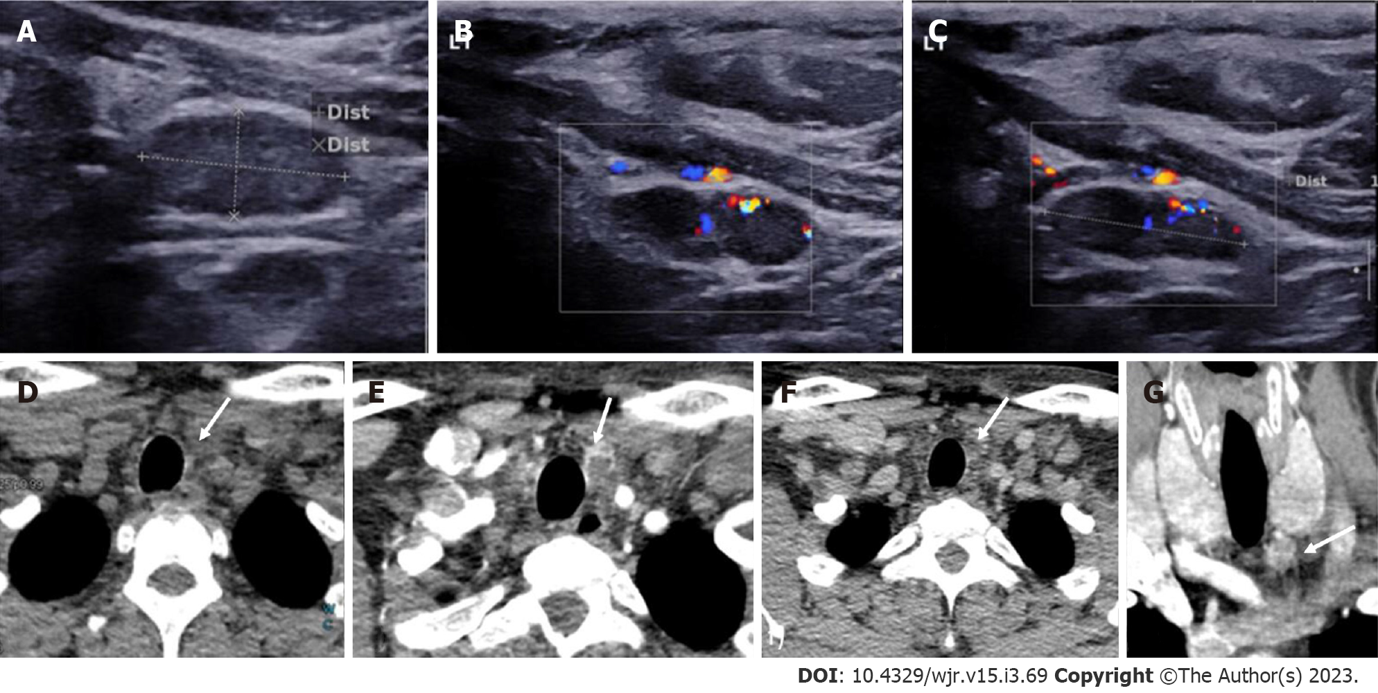
Figure 7 Left inferior parathyroid adenoma.
In a patient with raised parathormone levels (290 IU), grey scale ultrasound A: and colour doppler flow imaging; B and C: Showed a hypoechoic lesion with vascularity just below the left lobe of the; D: 4-dimensional computed tomography showed the lesion to be hypodense on noncontrast computer tomography; E: Hyperenhancing with central necrosis on arterial phase; F: Washout on the venous phase; G: Coronal image better demonstrates the lesion.
- Citation: Gulati S, Chumber S, Puri G, Spalkit S, Damle NA, Das CJ. Multi-modality parathyroid imaging: A shifting paradigm. World J Radiol 2023; 15(3): 69-82
- URL: https://www.wjgnet.com/1949-8470/full/v15/i3/69.htm
- DOI: https://dx.doi.org/10.4329/wjr.v15.i3.69


