Copyright
©The Author(s) 2023.
World J Radiol. Mar 28, 2023; 15(3): 69-82
Published online Mar 28, 2023. doi: 10.4329/wjr.v15.i3.69
Published online Mar 28, 2023. doi: 10.4329/wjr.v15.i3.69
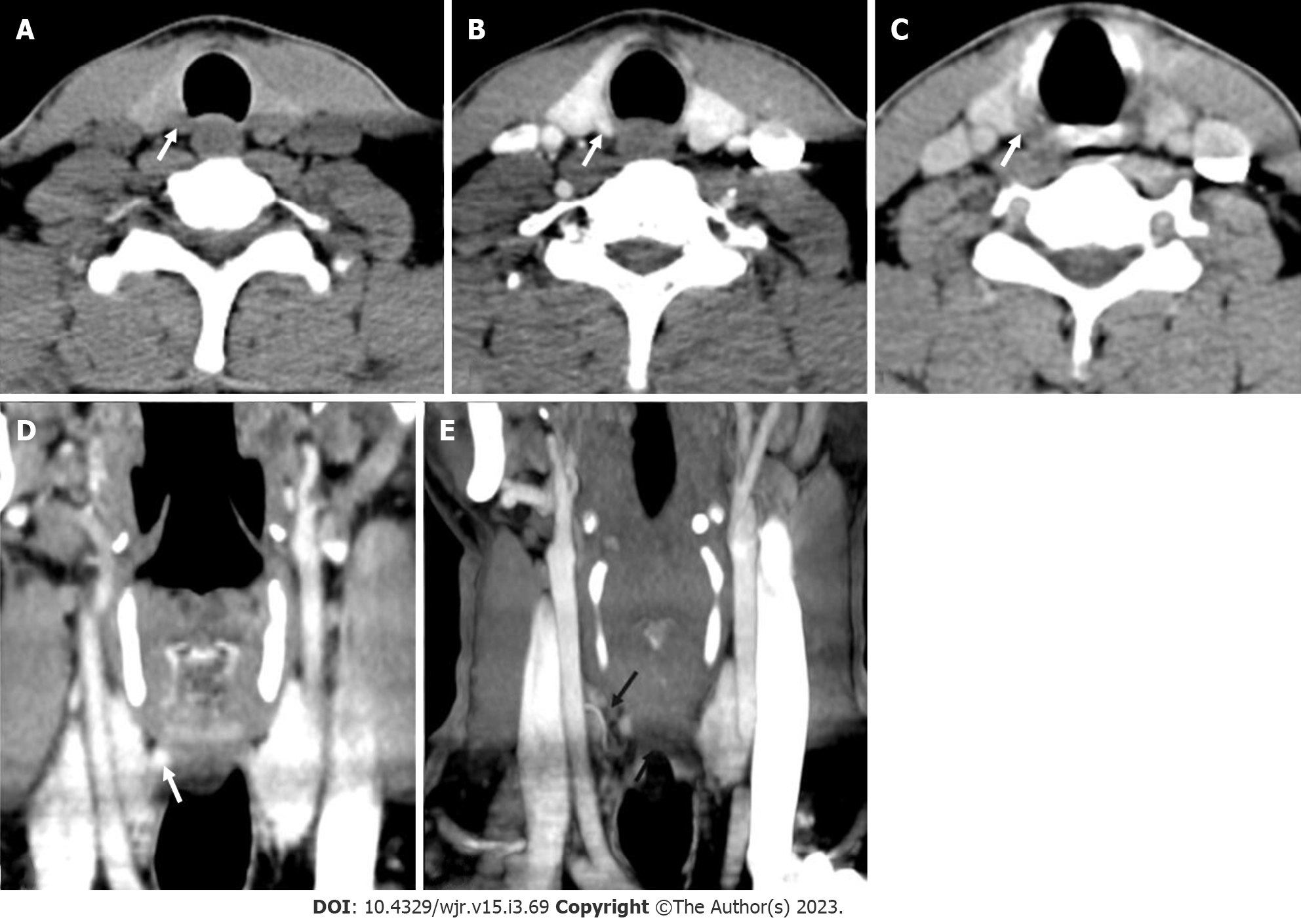
Figure 6 Right superior adenoma: Four-dimensional computed tomography.
A: Non-contrast computed tomography shows a small oval hypodense lesion which shows B: Intense enhancement on the arterial phase; C: Washout on the venous phase consistent with right superior parathyroid adenoma; D: Coronal image and E: Coronal maximum intensity projection image in the arterial phase better demonstrate the lesion with the feeding vessel (black arrow).
- Citation: Gulati S, Chumber S, Puri G, Spalkit S, Damle NA, Das CJ. Multi-modality parathyroid imaging: A shifting paradigm. World J Radiol 2023; 15(3): 69-82
- URL: https://www.wjgnet.com/1949-8470/full/v15/i3/69.htm
- DOI: https://dx.doi.org/10.4329/wjr.v15.i3.69


