Copyright
©The Author(s) 2023.
World J Radiol. Mar 28, 2023; 15(3): 56-68
Published online Mar 28, 2023. doi: 10.4329/wjr.v15.i3.56
Published online Mar 28, 2023. doi: 10.4329/wjr.v15.i3.56
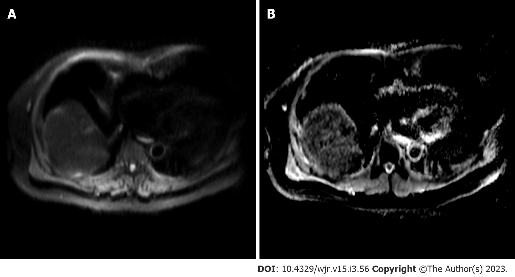
Figure 6 Diffusion-weighted imaging.
A: Diffusion-weighted imaging sequence; B: Apparent diffusion coefficient (ADC) map. Increased signal can be observed within the segment VIII lesion with corresponding hypointensity on ADC map, compatible with diffusion restriction.
- Citation: Criss C, Nagar AM, Makary MS. Hepatocellular carcinoma: State of the art diagnostic imaging. World J Radiol 2023; 15(3): 56-68
- URL: https://www.wjgnet.com/1949-8470/full/v15/i3/56.htm
- DOI: https://dx.doi.org/10.4329/wjr.v15.i3.56


