Copyright
©The Author(s) 2023.
World J Radiol. Feb 28, 2023; 15(2): 42-55
Published online Feb 28, 2023. doi: 10.4329/wjr.v15.i2.42
Published online Feb 28, 2023. doi: 10.4329/wjr.v15.i2.42
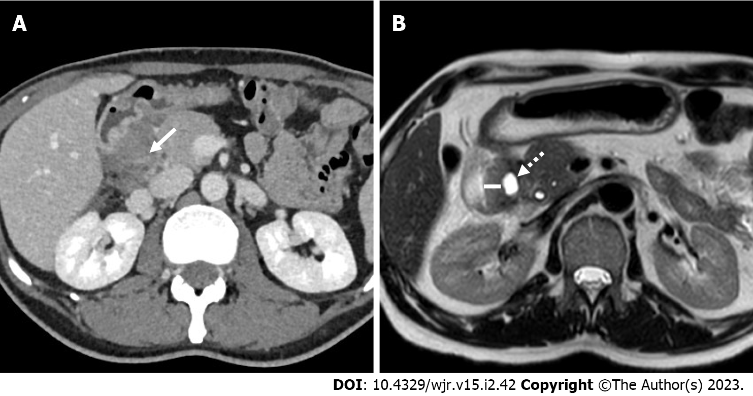
Figure 3 A 49-year-old male patient with weight loss and abdominal pain.
A: Axial 3 mm thick multiplanar reconstruction of portal venous phase computed tomography acquisition shows a hypodense mass in the groove region with patchy enhancement (arrow); B: Axial T2-weighted magnetic resonance imaging image acquired 2 mo later clearly shows eccentric second duodenal portion wall thickening (line) with cystic component (dotted arrow).
- Citation: Bonatti M, De Pretis N, Zamboni GA, Brillo A, Crinò SF, Valletta R, Lombardo F, Mansueto G, Frulloni L. Imaging of paraduodenal pancreatitis: A systematic review. World J Radiol 2023; 15(2): 42-55
- URL: https://www.wjgnet.com/1949-8470/full/v15/i2/42.htm
- DOI: https://dx.doi.org/10.4329/wjr.v15.i2.42


