Copyright
©The Author(s) 2023.
World J Radiol. Oct 28, 2023; 15(10): 281-292
Published online Oct 28, 2023. doi: 10.4329/wjr.v15.i10.281
Published online Oct 28, 2023. doi: 10.4329/wjr.v15.i10.281
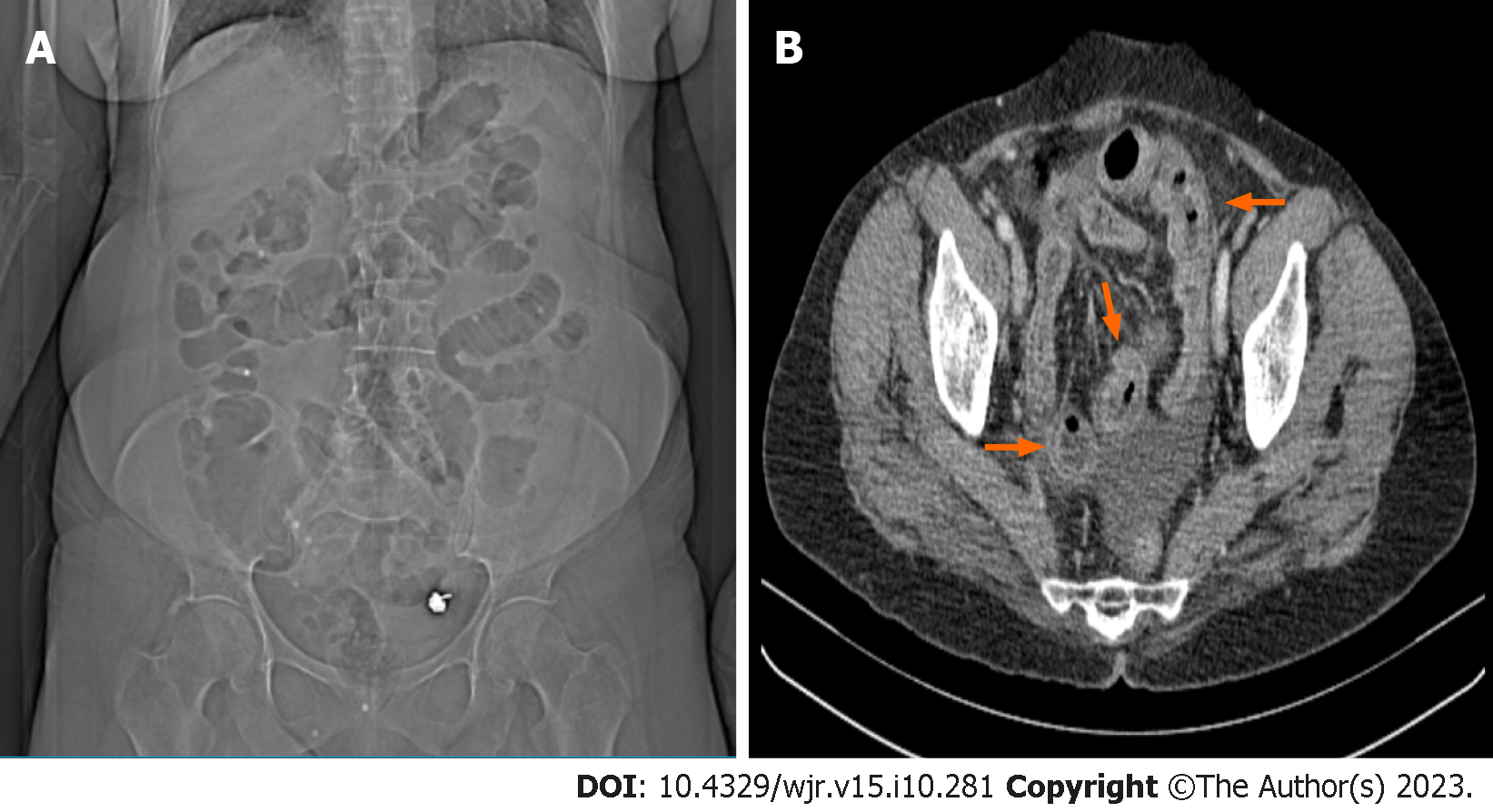
Figure 4 A 61-year-old female patient admitted to the emergency department complaining of abdominal pain.
A: Radiolucent air values in the large and small intestine segments are seen in the computed tomography (CT) scenogram image; B: Axial CT image shows trilaminar thickness increase (red arrows) in the small intestine wall compatible with ileitis, dilated appearance in the mesenteric vascular structures and free fluid in the pelvic area. Following observation of the scenogram images, the first observer reported a possible obstruction at the small intestine level, the second observer reported a possible obstruction at the large intestine level, and the third observer reported that no mechanical obstruction was detected.
- Citation: Kadirhan O, Kızılgoz V, Aydin S, Bilici E, Bayat E, Kantarci M. Does the use of computed tomography scenogram alone enable diagnosis in cases of bowel obstruction? World J Radiol 2023; 15(10): 281-292
- URL: https://www.wjgnet.com/1949-8470/full/v15/i10/281.htm
- DOI: https://dx.doi.org/10.4329/wjr.v15.i10.281


