Copyright
©The Author(s) 2023.
World J Radiol. Oct 28, 2023; 15(10): 281-292
Published online Oct 28, 2023. doi: 10.4329/wjr.v15.i10.281
Published online Oct 28, 2023. doi: 10.4329/wjr.v15.i10.281
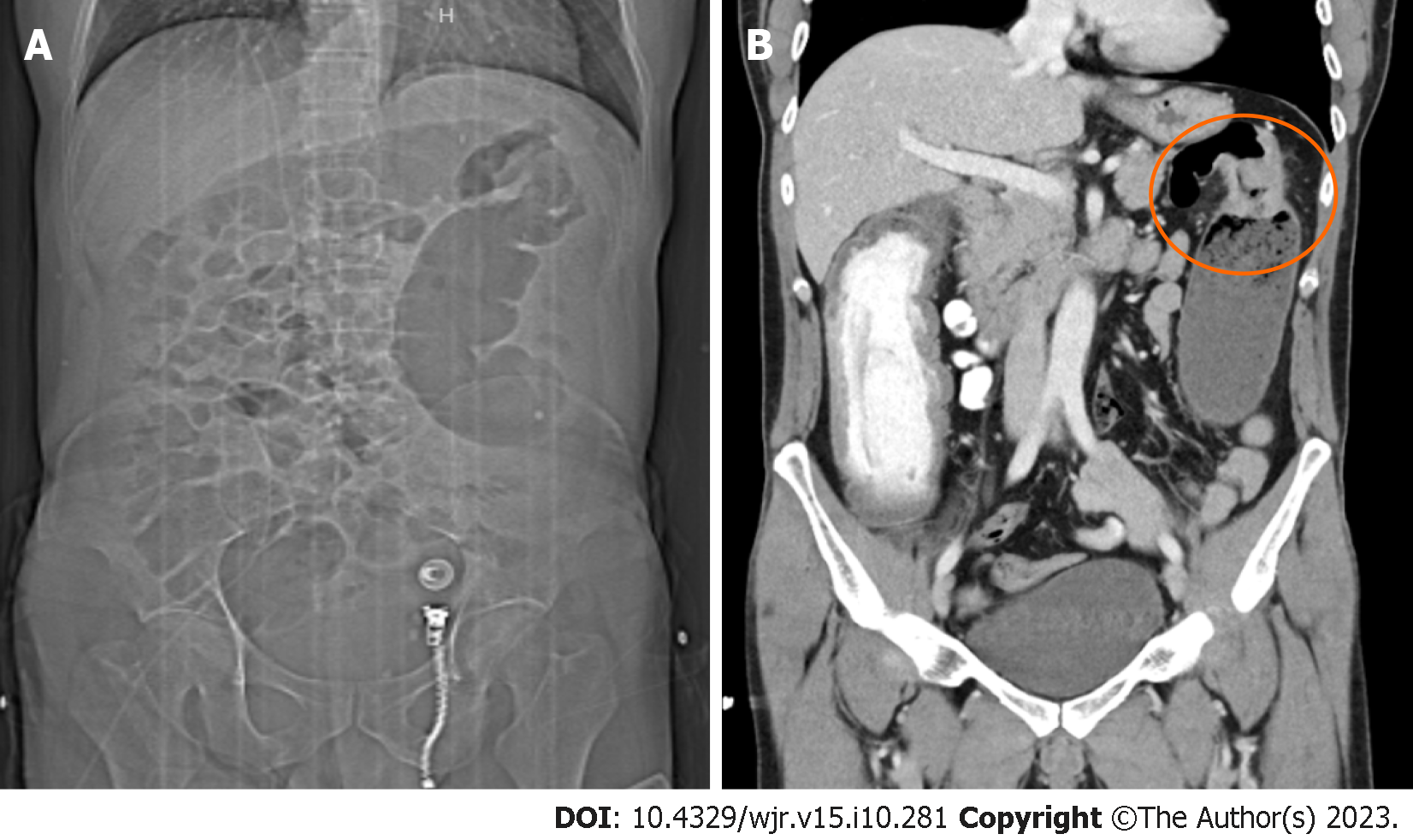
Figure 2 A 49-year-old male patient admitted to the emergency room with complaints of abdominal pain and vomiting.
A: In the computed tomography (CT) scenogram, sudden narrowing at the level of the splenic flexure and distension in the proximal and distal colon loops are seen; B: In the coronal CT image, a mass lesion causing a circular increase in wall thickness is seen at the level of the splenic flexure. All three observers reported a possible obstruction at the level of the large intestine.
- Citation: Kadirhan O, Kızılgoz V, Aydin S, Bilici E, Bayat E, Kantarci M. Does the use of computed tomography scenogram alone enable diagnosis in cases of bowel obstruction? World J Radiol 2023; 15(10): 281-292
- URL: https://www.wjgnet.com/1949-8470/full/v15/i10/281.htm
- DOI: https://dx.doi.org/10.4329/wjr.v15.i10.281


