Copyright
©The Author(s) 2022.
World J Radiol. Sep 28, 2022; 14(9): 342-351
Published online Sep 28, 2022. doi: 10.4329/wjr.v14.i9.342
Published online Sep 28, 2022. doi: 10.4329/wjr.v14.i9.342
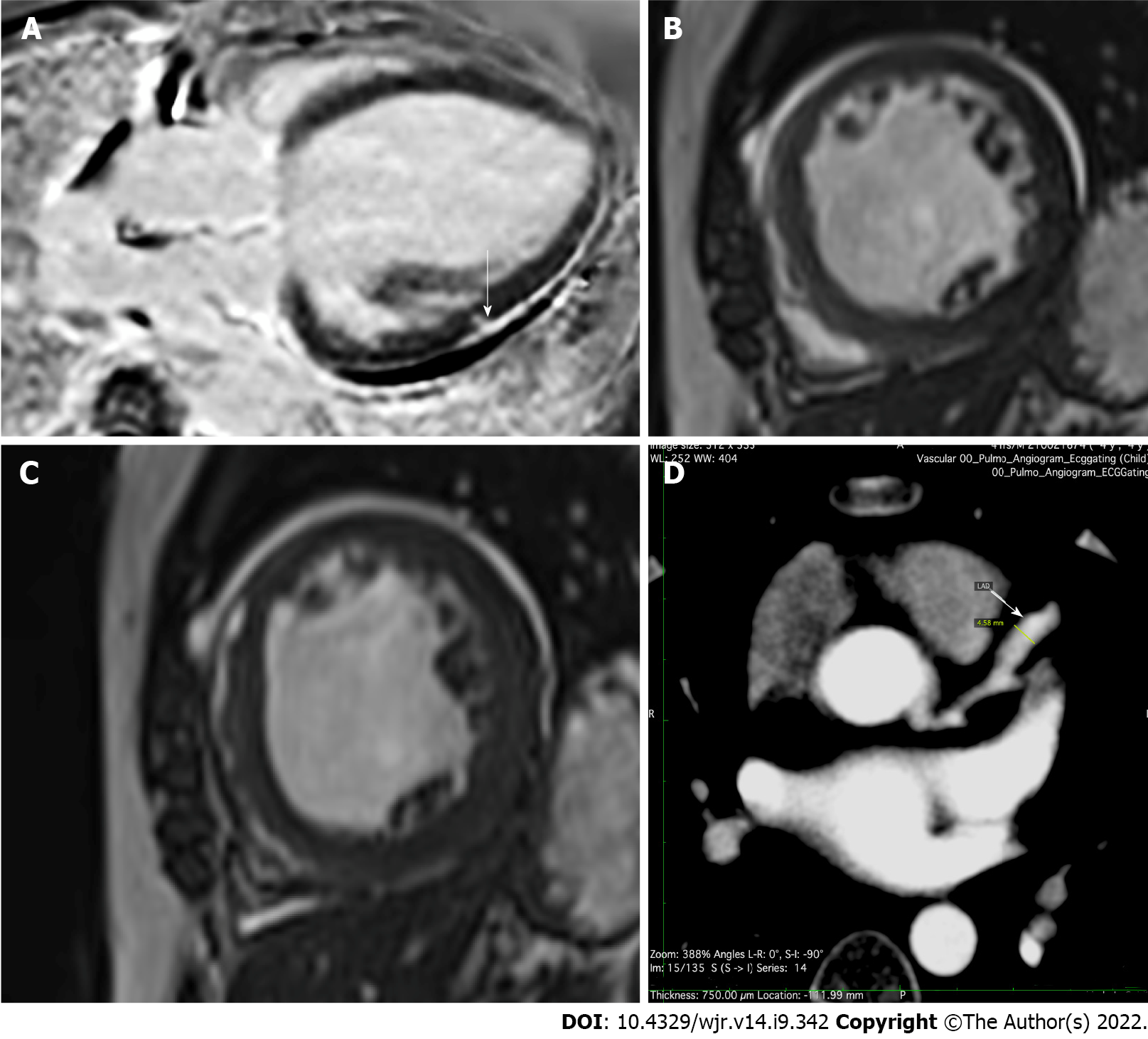
Figure 1 Post coronavirus disease 2019 imaging.
A: Myocarditis: Magnetic resonance late gadolinium enhancement imaging, 4 chamber view. Subepicardial scar with focal myocardial extension (arrow) in the mid anterolateral segment of the left ventricle; B and C: Dilated cardiomyopathy: Bright blood T2 weighted cine imaging in short-axis 2 chamber view showing a dilated left ventricle. Patient had a history of coronavirus disease 2019 (COVID-19) infection a year ago followed by increasing dyspnea. Magnetic resonance imaging revealed severe left ventricular dysfunction and asynchronous left ventricle contractions, B: End diastole; C: End systole; D: Coronary artery aneurysm: Computed tomography angiography in a 4-year-old child reveals a fusiform aneurysm of the left anterior descending coronary artery (arrow). The patient had a history of COVID-19 8 mo ago and was following up for the same.
- Citation: Merchant SA, Nadkarni P, Shaikh MJS. Augmentation of literature review of COVID-19 radiology. World J Radiol 2022; 14(9): 342-351
- URL: https://www.wjgnet.com/1949-8470/full/v14/i9/342.htm
- DOI: https://dx.doi.org/10.4329/wjr.v14.i9.342


