Copyright
©The Author(s) 2022.
World J Radiol. Sep 28, 2022; 14(9): 329-341
Published online Sep 28, 2022. doi: 10.4329/wjr.v14.i9.329
Published online Sep 28, 2022. doi: 10.4329/wjr.v14.i9.329
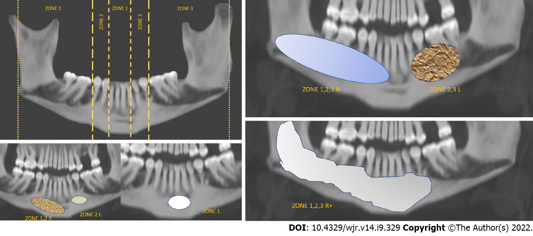
Figure 1 Location of every lesion was classified into the following zones: 1, limited to the incisors; 2, limited to the canine and premolars; and 3, limited to the molars and posterior mandible.
A similar classification was applied to the maxilla. Lesions extending over multiple zones were classified as such, and a suffix of R or L was used to denote right or left-sided location. When the lesion crossed the midline across multiple zones, + was used to denote the same.
- Citation: Ghosh A, Lakshmanan M, Manchanda S, Bhalla AS, Kumar P, Bhutia O, Mridha AR. Contrast-enhanced multidetector computed tomography features and histogram analysis can differentiate ameloblastomas from central giant cell granulomas . World J Radiol 2022; 14(9): 329-341
- URL: https://www.wjgnet.com/1949-8470/full/v14/i9/329.htm
- DOI: https://dx.doi.org/10.4329/wjr.v14.i9.329


