Copyright
©The Author(s) 2022.
World J Radiol. Sep 28, 2022; 14(9): 319-328
Published online Sep 28, 2022. doi: 10.4329/wjr.v14.i9.319
Published online Sep 28, 2022. doi: 10.4329/wjr.v14.i9.319
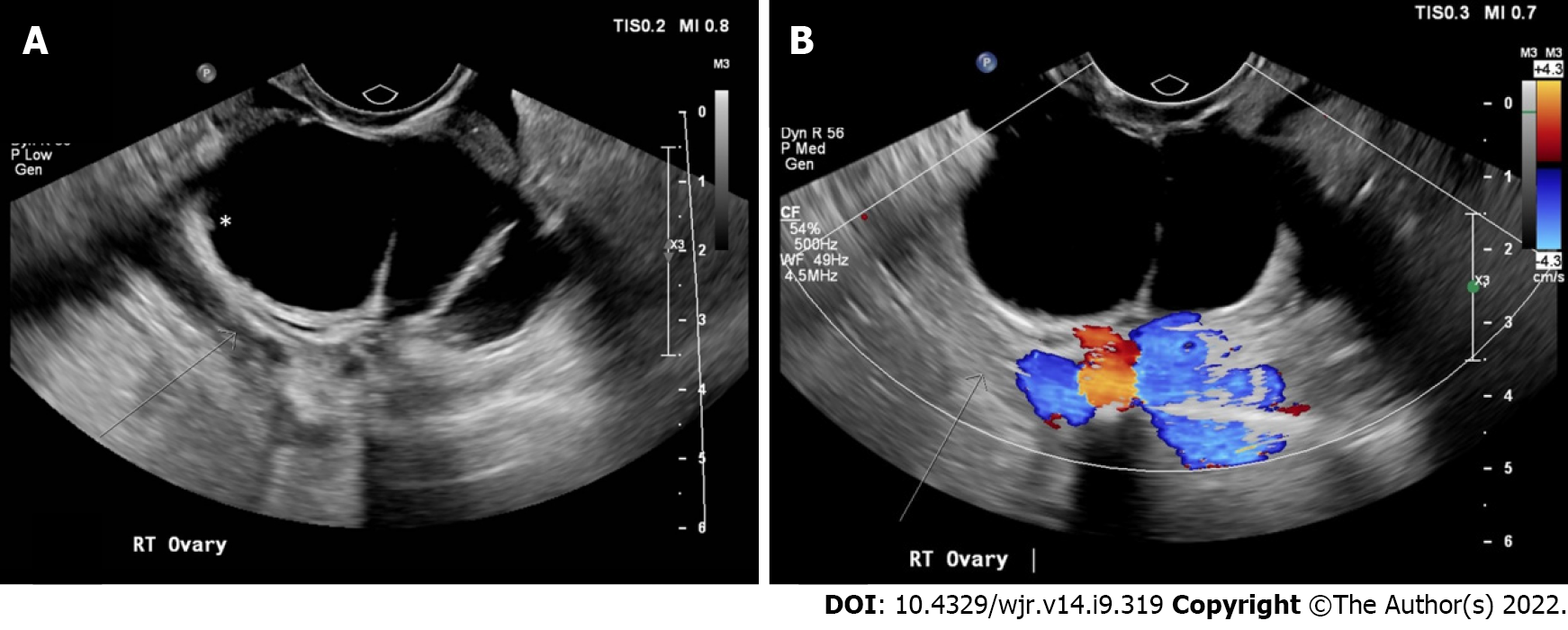
Figure 3 An example of a right ovarian cystic lesion misclassified as a “multilocular cyst < 10 cm, smooth inner wall, color score 1-3” (Ovarian-Adnexal Reporting and Data System 3).
A: Static gray-scale images; B: Static color Doppler ultrasound images. Static gray-scale and color Doppler ultrasound images show a multilocular cyst with a subtle non-uniform (irregular) inner wall with solid components < 3 mm in height (white asterisk) (ovarian-adnexal reporting and data system 4) (2).
- Citation: Katlariwala P, Wilson MP, Pi Y, Chahal BS, Croutze R, Patel D, Patel V, Low G. Reliability of ultrasound ovarian-adnexal reporting and data system amongst less experienced readers before and after training. World J Radiol 2022; 14(9): 319-328
- URL: https://www.wjgnet.com/1949-8470/full/v14/i9/319.htm
- DOI: https://dx.doi.org/10.4329/wjr.v14.i9.319


