Copyright
©The Author(s) 2022.
World J Radiol. Aug 28, 2022; 14(8): 272-285
Published online Aug 28, 2022. doi: 10.4329/wjr.v14.i8.272
Published online Aug 28, 2022. doi: 10.4329/wjr.v14.i8.272
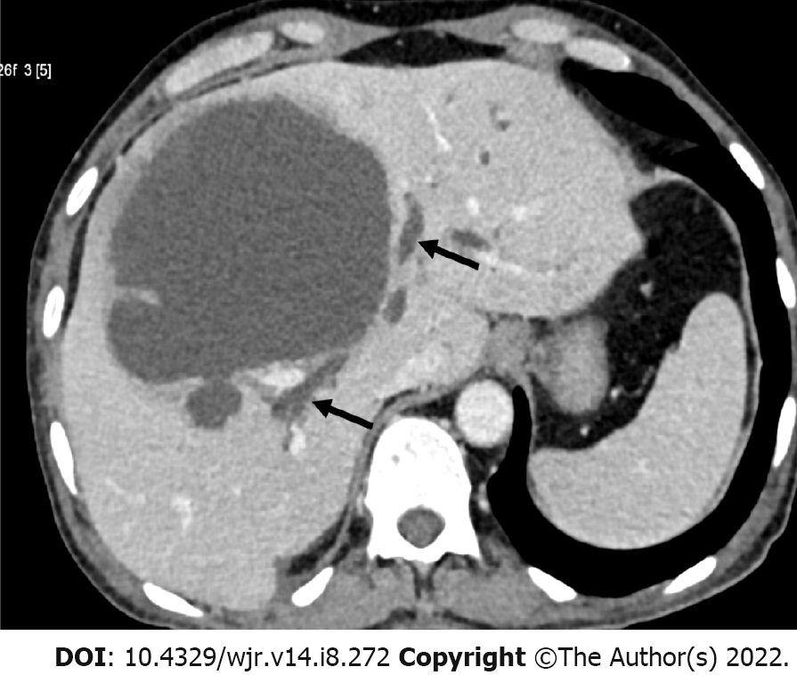
Figure 4 Axial computed tomography of a 60-year-old man showing a large abscess in segment IV of the liver near the porta hepatis.
Note the duct dilation (arrows) that resulted from rupture of the abscess into the central bile ducts. He was managed with catheter drainage. Bilious fluid draining through the catheter was observed for several weeks in this patient.
- Citation: Priyadarshi RN, Kumar R, Anand U. Amebic liver abscess: Clinico-radiological findings and interventional management. World J Radiol 2022; 14(8): 272-285
- URL: https://www.wjgnet.com/1949-8470/full/v14/i8/272.htm
- DOI: https://dx.doi.org/10.4329/wjr.v14.i8.272


