Copyright
©The Author(s) 2022.
World J Radiol. Aug 28, 2022; 14(8): 272-285
Published online Aug 28, 2022. doi: 10.4329/wjr.v14.i8.272
Published online Aug 28, 2022. doi: 10.4329/wjr.v14.i8.272
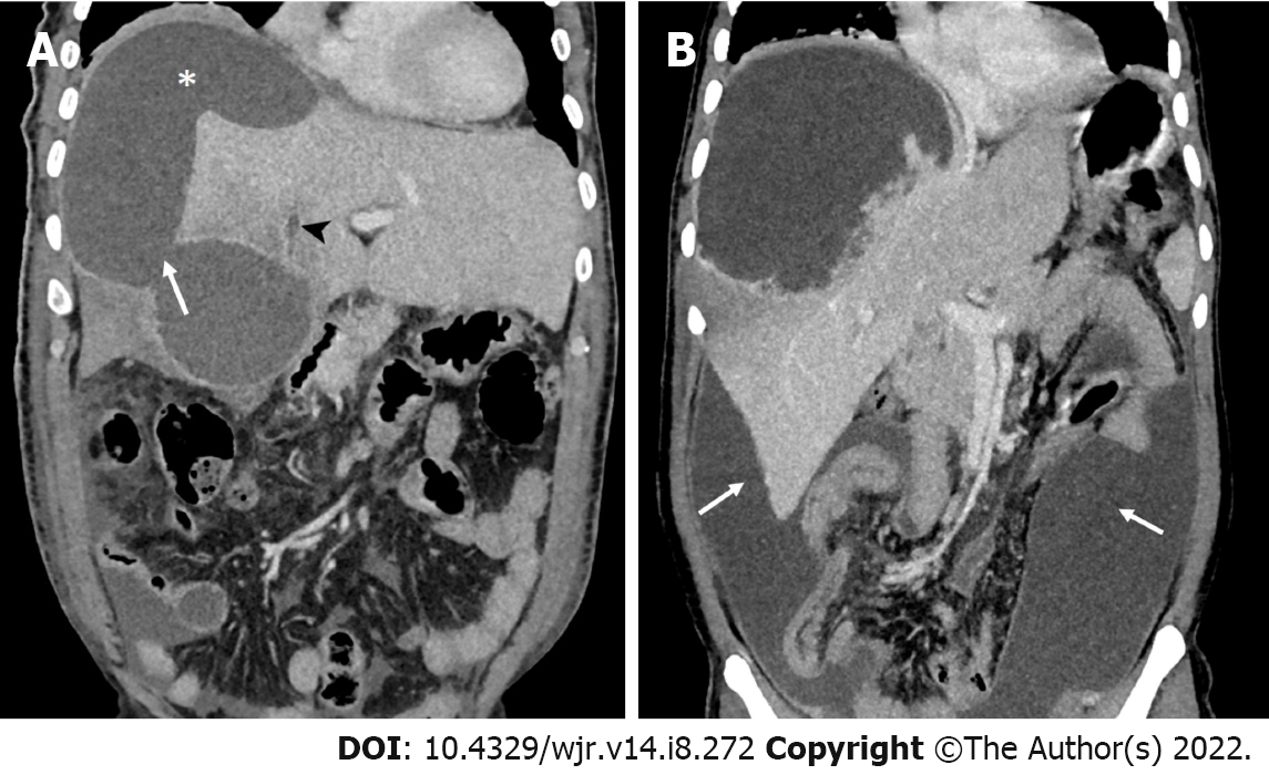
Figure 3 Computed tomography image.
A: Computed tomography image (coronal view) demonstrating a contained rupture. A fluid collection that is localized in the subphrenic space (asterisk). Note the wide rent in the abscess (arrow). Additional imaging features of an aggressive disease in this image are the presence of ascites and thrombus in a segment of the hepatic vein (arrowhead); B: Free intraperitoneal rupture in a 40-year-old man who presented with features of generalized peritonitis. Coronal computed tomography image showing a large amebic abscess with an irregular edge in the right lobe and diffuse intraperitoneal fluid collection (arrows).
- Citation: Priyadarshi RN, Kumar R, Anand U. Amebic liver abscess: Clinico-radiological findings and interventional management. World J Radiol 2022; 14(8): 272-285
- URL: https://www.wjgnet.com/1949-8470/full/v14/i8/272.htm
- DOI: https://dx.doi.org/10.4329/wjr.v14.i8.272


