Copyright
©The Author(s) 2022.
World J Radiol. Aug 28, 2022; 14(8): 272-285
Published online Aug 28, 2022. doi: 10.4329/wjr.v14.i8.272
Published online Aug 28, 2022. doi: 10.4329/wjr.v14.i8.272
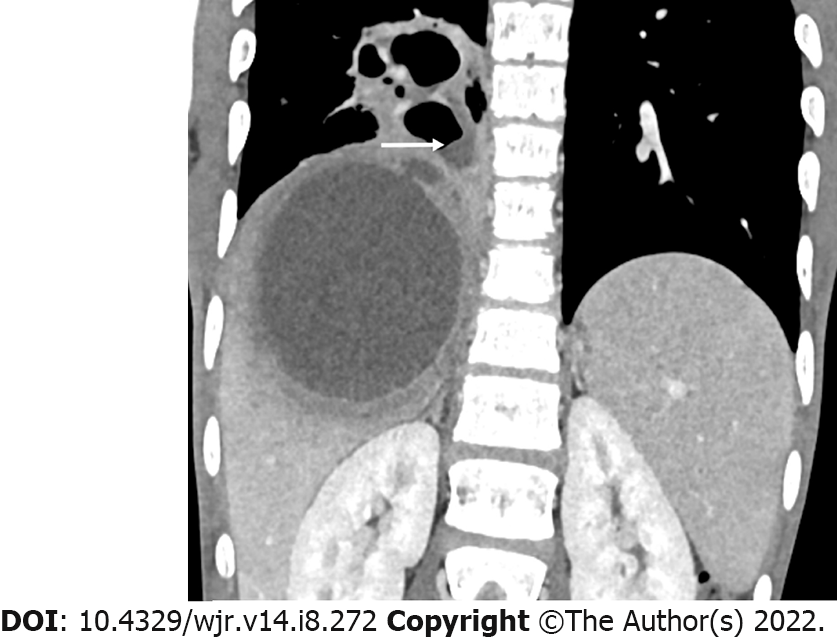
Figure 2 Computed tomography image (coronal view) of a patient who presented with productive cough and mild upper abdominal pain for more than four weeks.
Note the rupture of a subdiaphragmatic abscess into the lung resulting in the formation of a lung abscess. The air-fluid level in the lung abscess (arrow) indicates fistulous communication between the lung abscess and the bronchus.
- Citation: Priyadarshi RN, Kumar R, Anand U. Amebic liver abscess: Clinico-radiological findings and interventional management. World J Radiol 2022; 14(8): 272-285
- URL: https://www.wjgnet.com/1949-8470/full/v14/i8/272.htm
- DOI: https://dx.doi.org/10.4329/wjr.v14.i8.272


