Copyright
©The Author(s) 2022.
World J Radiol. Aug 28, 2022; 14(8): 272-285
Published online Aug 28, 2022. doi: 10.4329/wjr.v14.i8.272
Published online Aug 28, 2022. doi: 10.4329/wjr.v14.i8.272
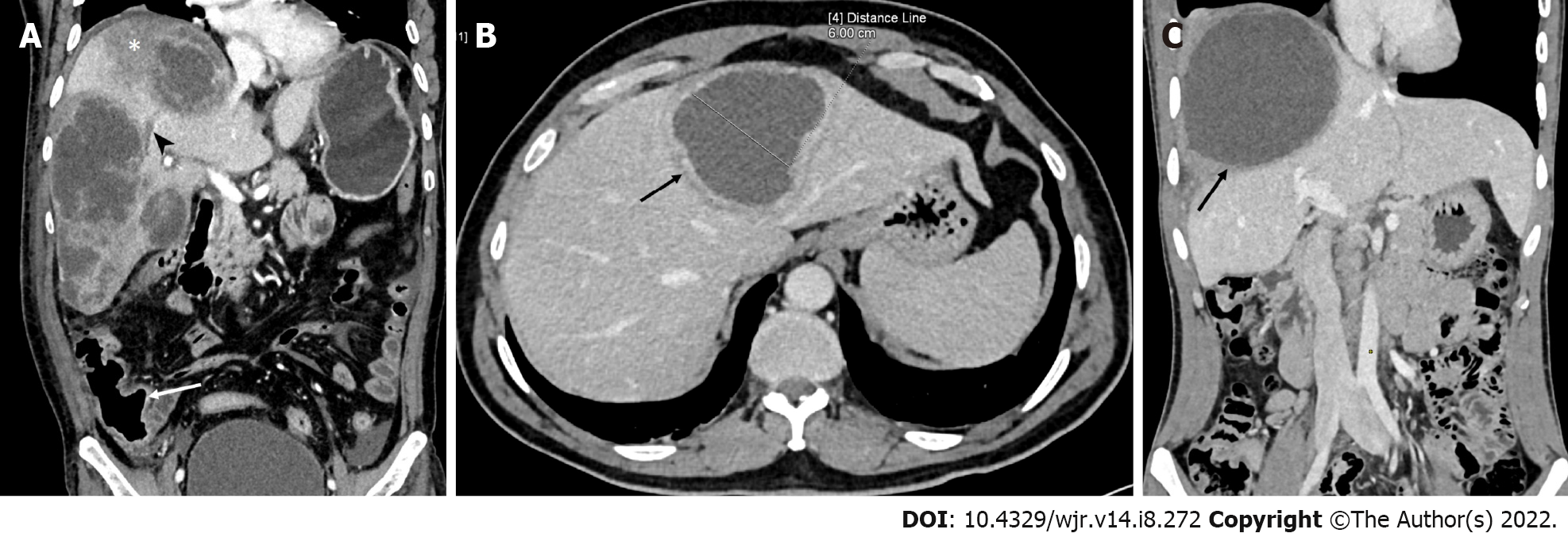
Figure 1 Computed tomography images.
A: Computed tomography (CT) (coronal image) demonstrating the characteristic imaging findings of an acute aggressive abscess (type I pattern) in a 60-year-old man who presented with sepsis-like features and markedly deranged laboratory parameters. There are multiple abscesses in the right lobe with irregular ragged edges, multiple septa and heterogeneous densities indicating partially liquefied tissue. Also, note the presence of a hypodense area in the surrounding parenchyma (asterisk) and right hepatic vein thrombosis (arrowhead). The thickened cecal wall (arrow) and mild ascites are also evident; B: CT of a typical case of subacute mild disease. The laboratory profile was near normal. The axial image shows an abscess in the left lobe with a well-defined wall showing rim enhancement (type II pattern). This patient presented with mild abdominal pain after 20 d of symptoms; C: CT image of a chronic indolent abscess (type III pattern). Coronal image of a 24-year-old man showing a large abscess with a thick non-enhancing wall in the right lobe. He had persistent pain in the right upper quadrant for two months despite complete resolution of fever and normalization of laboratory tests after metronidazole therapy.
- Citation: Priyadarshi RN, Kumar R, Anand U. Amebic liver abscess: Clinico-radiological findings and interventional management. World J Radiol 2022; 14(8): 272-285
- URL: https://www.wjgnet.com/1949-8470/full/v14/i8/272.htm
- DOI: https://dx.doi.org/10.4329/wjr.v14.i8.272


