Copyright
©The Author(s) 2022.
World J Radiol. Jul 28, 2022; 14(7): 209-218
Published online Jul 28, 2022. doi: 10.4329/wjr.v14.i7.209
Published online Jul 28, 2022. doi: 10.4329/wjr.v14.i7.209
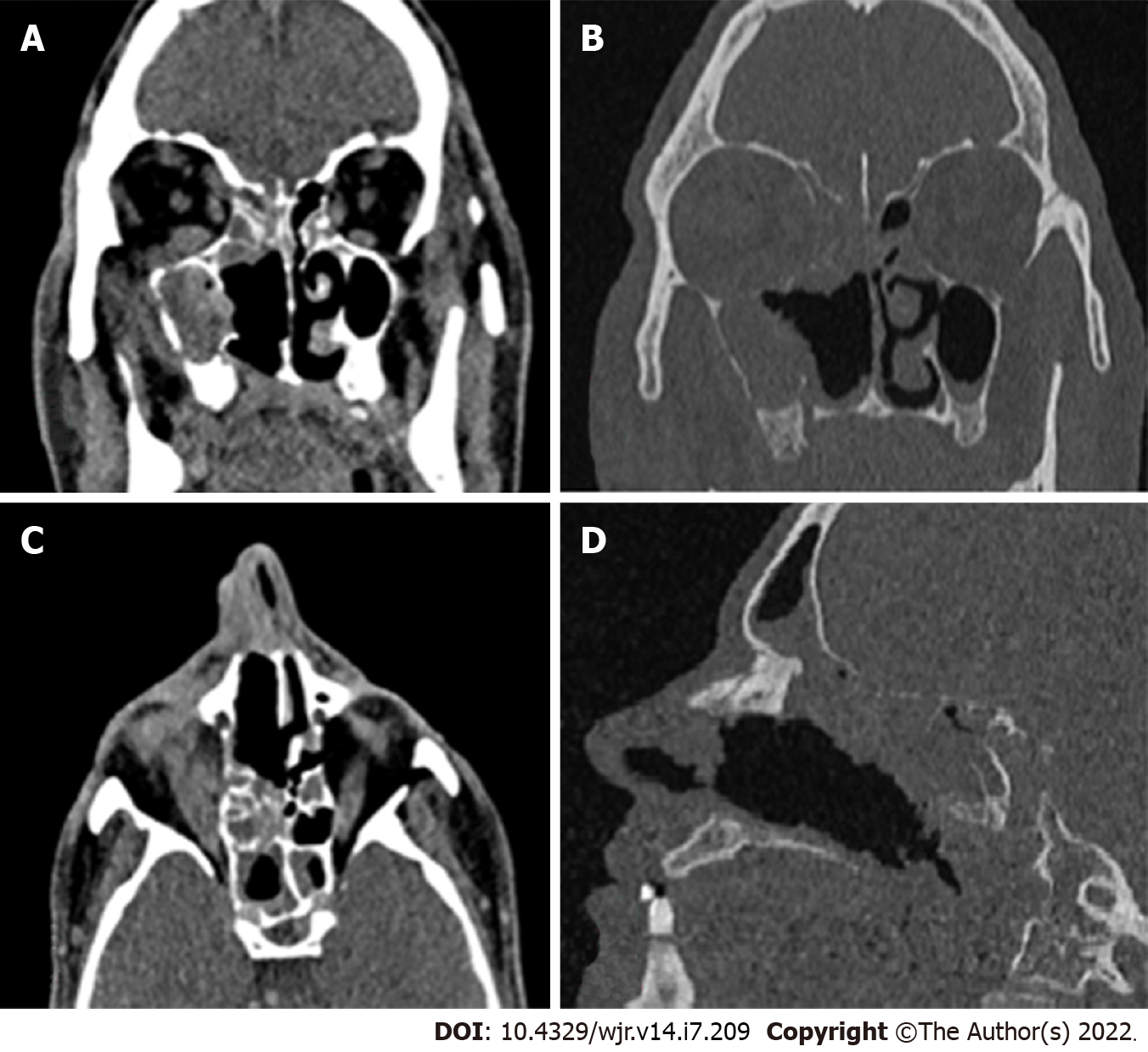
Figure 1 Non contrast computed tomography image of a 40 year old male post renal transplant patient showing.
A: Soft tissue density in maxillary, ethmoidal, sphenoidal and frontal sinus; B: Rarefaction of ethmoidal lamella, lamina papyracea and floor of anterior cranial fossa and erosion of maxillary walls; C: Section showing orbital involvement; D: Section showing erosions of the cribriform plate.
- Citation: Saneesh PS, Morampudi SC, Yelamanchi R. Radiological review of rhinocerebral mucormycosis cases during the COVID-19 Pandemic: A single-center experience. World J Radiol 2022; 14(7): 209-218
- URL: https://www.wjgnet.com/1949-8470/full/v14/i7/209.htm
- DOI: https://dx.doi.org/10.4329/wjr.v14.i7.209


