Copyright
©The Author(s) 2022.
World J Radiol. Jul 28, 2022; 14(7): 180-193
Published online Jul 28, 2022. doi: 10.4329/wjr.v14.i7.180
Published online Jul 28, 2022. doi: 10.4329/wjr.v14.i7.180
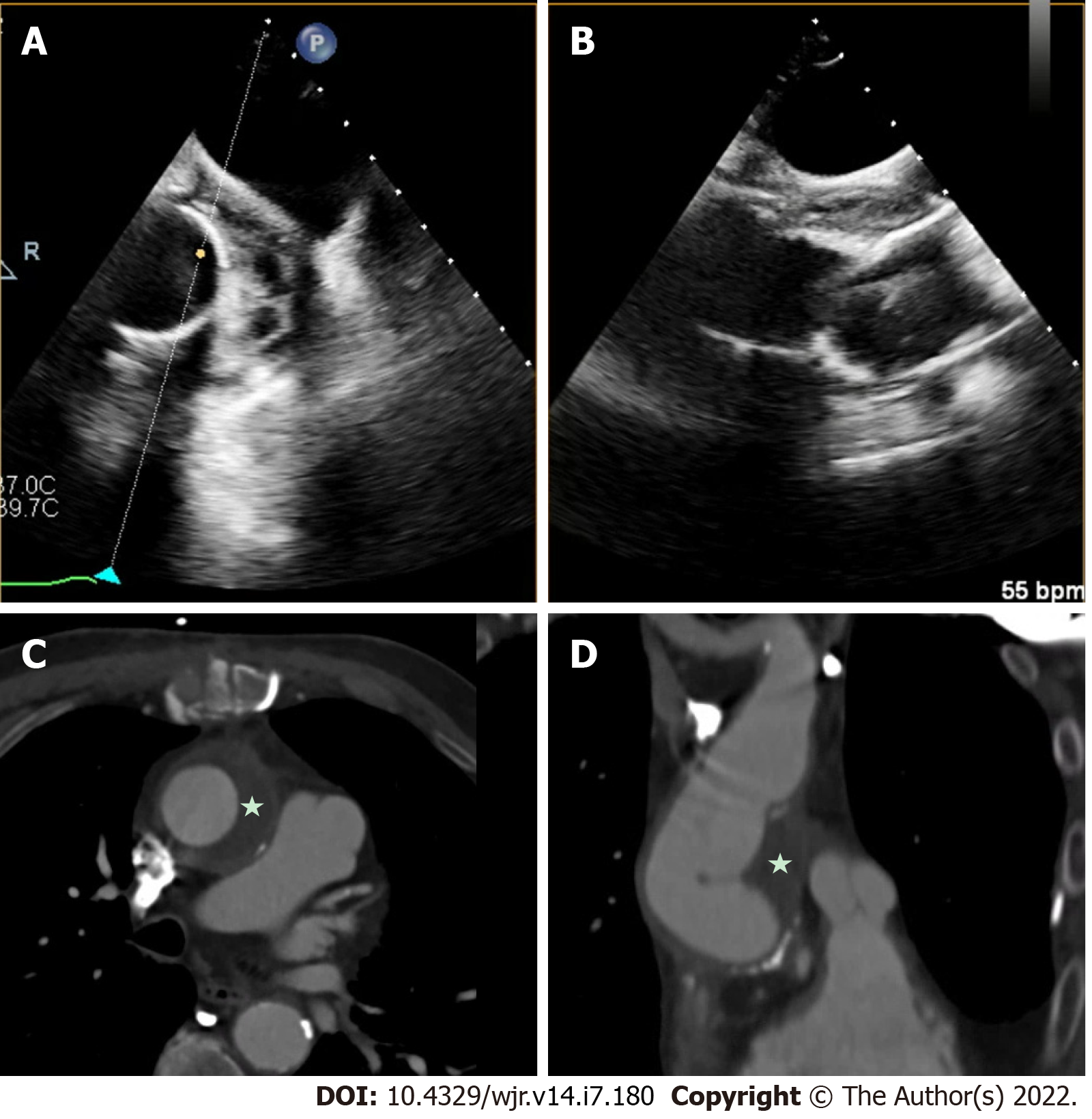
Figure 6 Aortic graft infection.
A and B: Infection of aortic grafts can be difficult to visualize on transesophageal echocardiography (TEE); C and D: This case demonstrates evidence of a peri-aortic graft echolucent space with stranding, which was challenging to image on TEE, and further characterization with cardiac computed tomography clearly demonstrated peri-aortic graft thickening (star) consistent with infection.
- Citation: Hughes D, Linchangco R, Reyaldeen R, Xu B. Expanding utility of cardiac computed tomography in infective endocarditis: A contemporary review . World J Radiol 2022; 14(7): 180-193
- URL: https://www.wjgnet.com/1949-8470/full/v14/i7/180.htm
- DOI: https://dx.doi.org/10.4329/wjr.v14.i7.180


