Copyright
©The Author(s) 2022.
World J Radiol. Jul 28, 2022; 14(7): 180-193
Published online Jul 28, 2022. doi: 10.4329/wjr.v14.i7.180
Published online Jul 28, 2022. doi: 10.4329/wjr.v14.i7.180
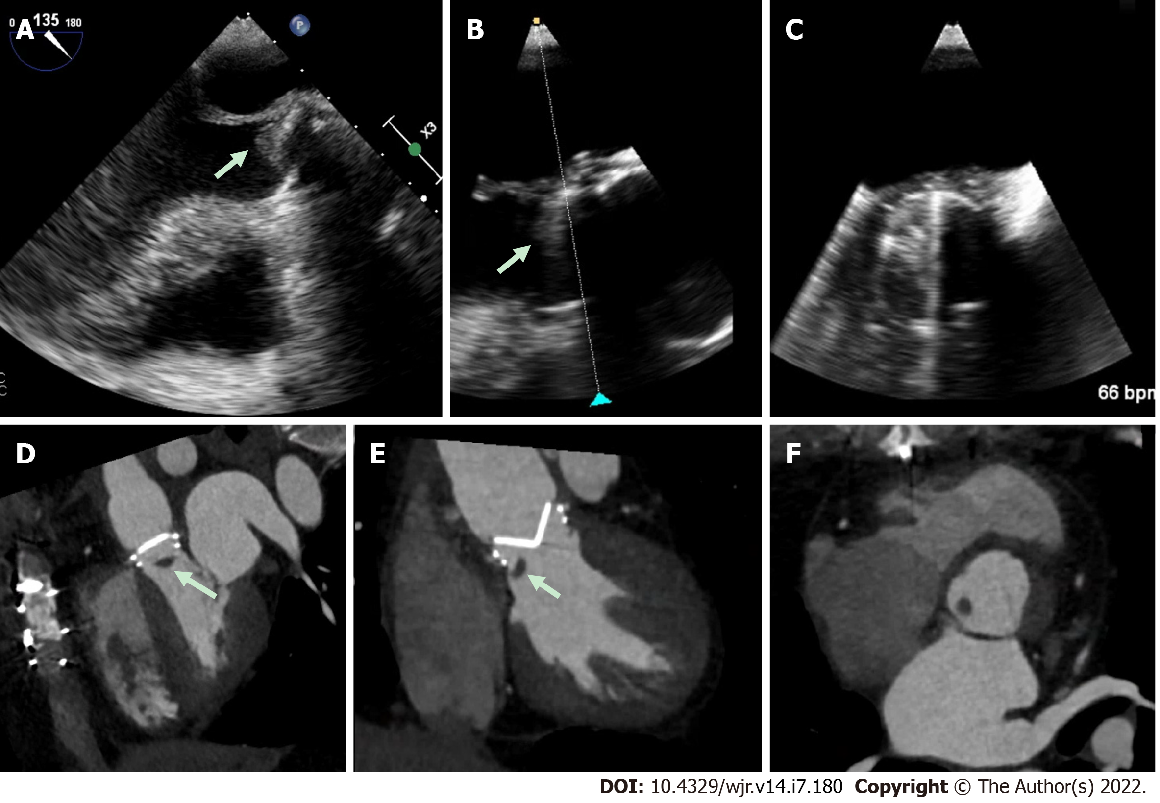
Figure 5 Prosthetic aortic valve with hypoattenuating lesion.
A-C: This patient presented with elevated prosthetic aortic valve gradients, and although transesophageal echocardiography imaging was suspicious for an echodensity (white arrow), imaging quality was challenging due to prosthetic metallic valve shadowing; D-F: Multi-phase 4-dimensional cardiac computed tomography was utilized which clearly demonstrated a hypoattenuating mass (white arrows) attached to the prosthetic valve. Despite suspicion for infection, intra-operative pathology revealed thrombus.
- Citation: Hughes D, Linchangco R, Reyaldeen R, Xu B. Expanding utility of cardiac computed tomography in infective endocarditis: A contemporary review . World J Radiol 2022; 14(7): 180-193
- URL: https://www.wjgnet.com/1949-8470/full/v14/i7/180.htm
- DOI: https://dx.doi.org/10.4329/wjr.v14.i7.180


