Copyright
©The Author(s) 2022.
World J Radiol. Jul 28, 2022; 14(7): 180-193
Published online Jul 28, 2022. doi: 10.4329/wjr.v14.i7.180
Published online Jul 28, 2022. doi: 10.4329/wjr.v14.i7.180
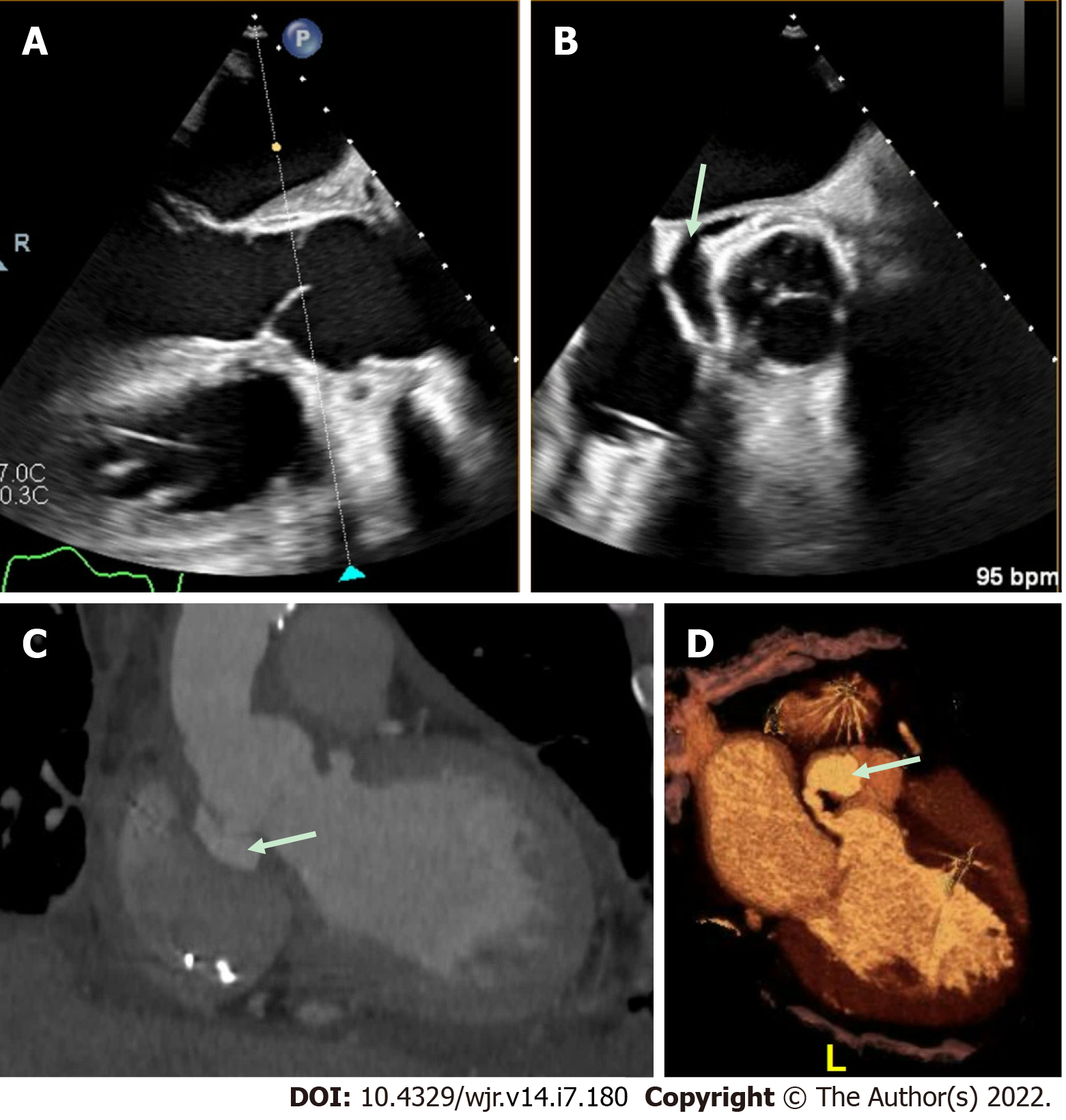
Figure 4 Aortic repair with left ventricular outflow tract pseudoaneurysm.
A and B: This patient had a complex aortic repair with a clear peri-valvular space (arrow) on transesophageal echocardiography (TEE); however, the exact origin was difficult to define by TEE imaging; C and D: Cardiac computed tomography demonstrated multiple pseudo-aneurysms arising from the left ventricular outflow tract (LVOT) with the largest, fistulous LVOT pseudo-aneurysm (arrow) best appreciated on 3-dimensional volume-rendering.
- Citation: Hughes D, Linchangco R, Reyaldeen R, Xu B. Expanding utility of cardiac computed tomography in infective endocarditis: A contemporary review . World J Radiol 2022; 14(7): 180-193
- URL: https://www.wjgnet.com/1949-8470/full/v14/i7/180.htm
- DOI: https://dx.doi.org/10.4329/wjr.v14.i7.180


