Copyright
©The Author(s) 2022.
World J Radiol. Jul 28, 2022; 14(7): 180-193
Published online Jul 28, 2022. doi: 10.4329/wjr.v14.i7.180
Published online Jul 28, 2022. doi: 10.4329/wjr.v14.i7.180
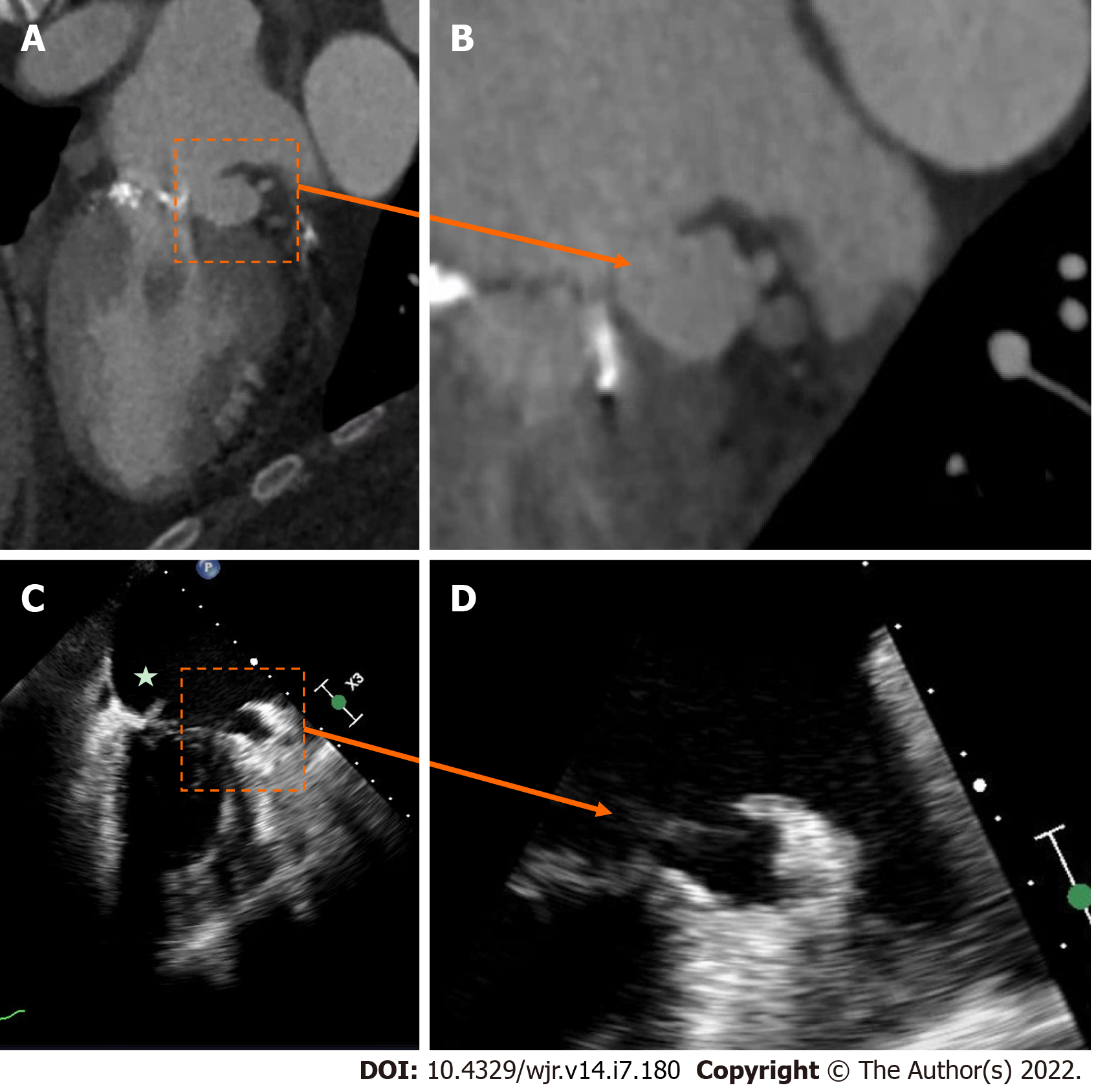
Figure 2 Native mitral valve infective endocarditis with pseudoaneurysm.
A and B: Mitral annular pseudo-aneurysm (dotted box) detected on cardiac computed tomography; C: Cardiac computed tomography with evidence of native mitral valve endocarditis on transesophageal echocardiography (star); D: Cardiac computed tomography with a prominent peri-annular cavity, consistent with a mitral annular pseudo-aneurysm.
- Citation: Hughes D, Linchangco R, Reyaldeen R, Xu B. Expanding utility of cardiac computed tomography in infective endocarditis: A contemporary review . World J Radiol 2022; 14(7): 180-193
- URL: https://www.wjgnet.com/1949-8470/full/v14/i7/180.htm
- DOI: https://dx.doi.org/10.4329/wjr.v14.i7.180


