Copyright
©The Author(s) 2022.
World J Radiol. Jun 28, 2022; 14(6): 165-176
Published online Jun 28, 2022. doi: 10.4329/wjr.v14.i6.165
Published online Jun 28, 2022. doi: 10.4329/wjr.v14.i6.165
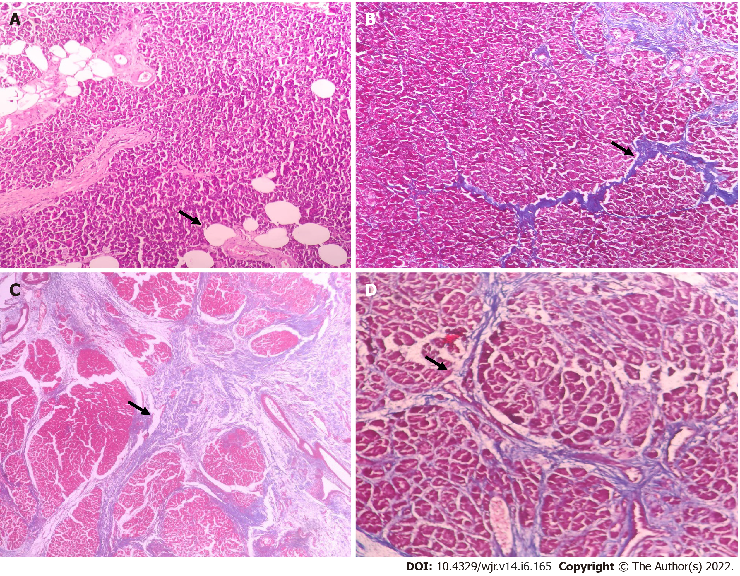
Figure 2 Histopathological evaluation of pancreatic neck fat fraction and fibrosis.
A: Photomicrograph showing moderate fat inclusion (hematoxylin and eosin [H&E], × 100); B: Photomicrograph showing heavy intralobular fibrosis (Masson's trichome stain, H&E, × 100); C: Photomicrograph showing heavy interlobular fibrosis (Masson's trichome stain, × 40); D: Photomicrograph showing weak intra and interlobular fibrosis (Masson's trichome stain, × 200).
- Citation: Gnanasekaran S, Durgesh S, Gurram R, Kalayarasan R, Pottakkat B, Rajeswari M, Srinivas BH, Ramesh A, Sahoo J. Do preoperative pancreatic computed tomography attenuation index and enhancement ratio predict pancreatic fistula after pancreaticoduodenectomy? World J Radiol 2022; 14(6): 165-176
- URL: https://www.wjgnet.com/1949-8470/full/v14/i6/165.htm
- DOI: https://dx.doi.org/10.4329/wjr.v14.i6.165


