Copyright
©The Author(s) 2022.
World J Radiol. Jun 28, 2022; 14(6): 155-164
Published online Jun 28, 2022. doi: 10.4329/wjr.v14.i6.155
Published online Jun 28, 2022. doi: 10.4329/wjr.v14.i6.155
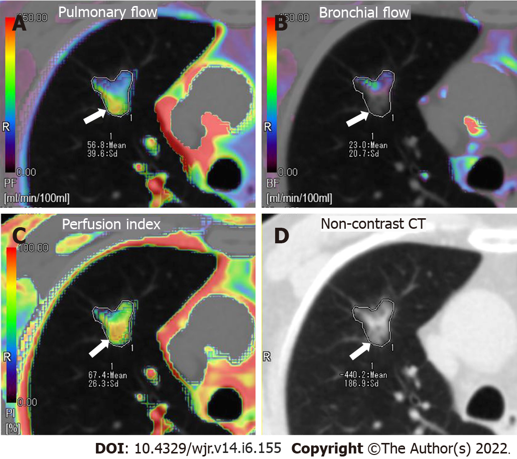
Figure 2 Axial colored perfusion maps in a 72-year-old male patient with mixed ground-glass nodule carcinoma located in the right superior lung.
The perfusion is heterogeneous throughout the lesion. Pulmonary flow (PF) is globally dominant, especially in the dorsal part of the lesion (arrow), which corresponds to the lower attenuation region of the mixed ground-glass nodule. A: Axial colored perfusion map of PF; B: Axial colored perfusion map of bronchial flow; C: Axial colored perfusion map of perfusion index; D: Axial non-contrast computed tomography.
- Citation: Wang C, Wu N, Zhang Z, Zhang LX, Yuan XD. Evaluation of the dual vascular supply patterns in ground-glass nodules with a dynamic volume computed tomography. World J Radiol 2022; 14(6): 155-164
- URL: https://www.wjgnet.com/1949-8470/full/v14/i6/155.htm
- DOI: https://dx.doi.org/10.4329/wjr.v14.i6.155


