Copyright
©The Author(s) 2022.
World J Radiol. Jun 28, 2022; 14(6): 137-150
Published online Jun 28, 2022. doi: 10.4329/wjr.v14.i6.137
Published online Jun 28, 2022. doi: 10.4329/wjr.v14.i6.137
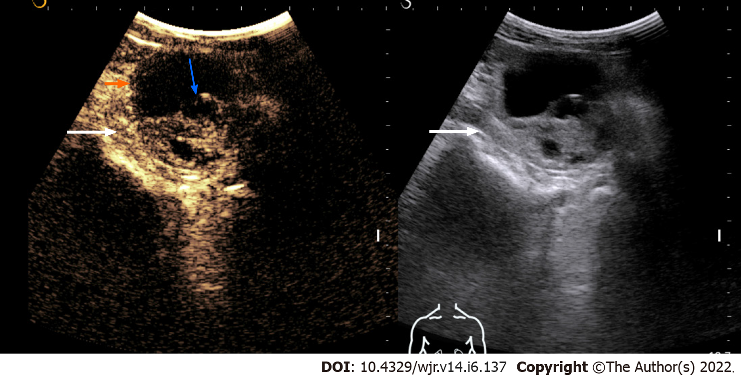
Figure 1 Contrast-enhanced ultrasonography images of a solid-cystic lesion in the left kidney showing thick nodular septal enhancement (blue arrow) and enhancement of solid component (long arrow).
Consistent with Bosniak category 4 lesion/malignant lesion. The lesion was resected and histology revealed clear cell renal cell carcinoma.
- Citation: Aggarwal A, Das CJ, Sharma S. Recent advances in imaging techniques of renal masses. World J Radiol 2022; 14(6): 137-150
- URL: https://www.wjgnet.com/1949-8470/full/v14/i6/137.htm
- DOI: https://dx.doi.org/10.4329/wjr.v14.i6.137


