Copyright
©The Author(s) 2022.
World J Radiol. Jun 28, 2022; 14(6): 114-136
Published online Jun 28, 2022. doi: 10.4329/wjr.v14.i6.114
Published online Jun 28, 2022. doi: 10.4329/wjr.v14.i6.114
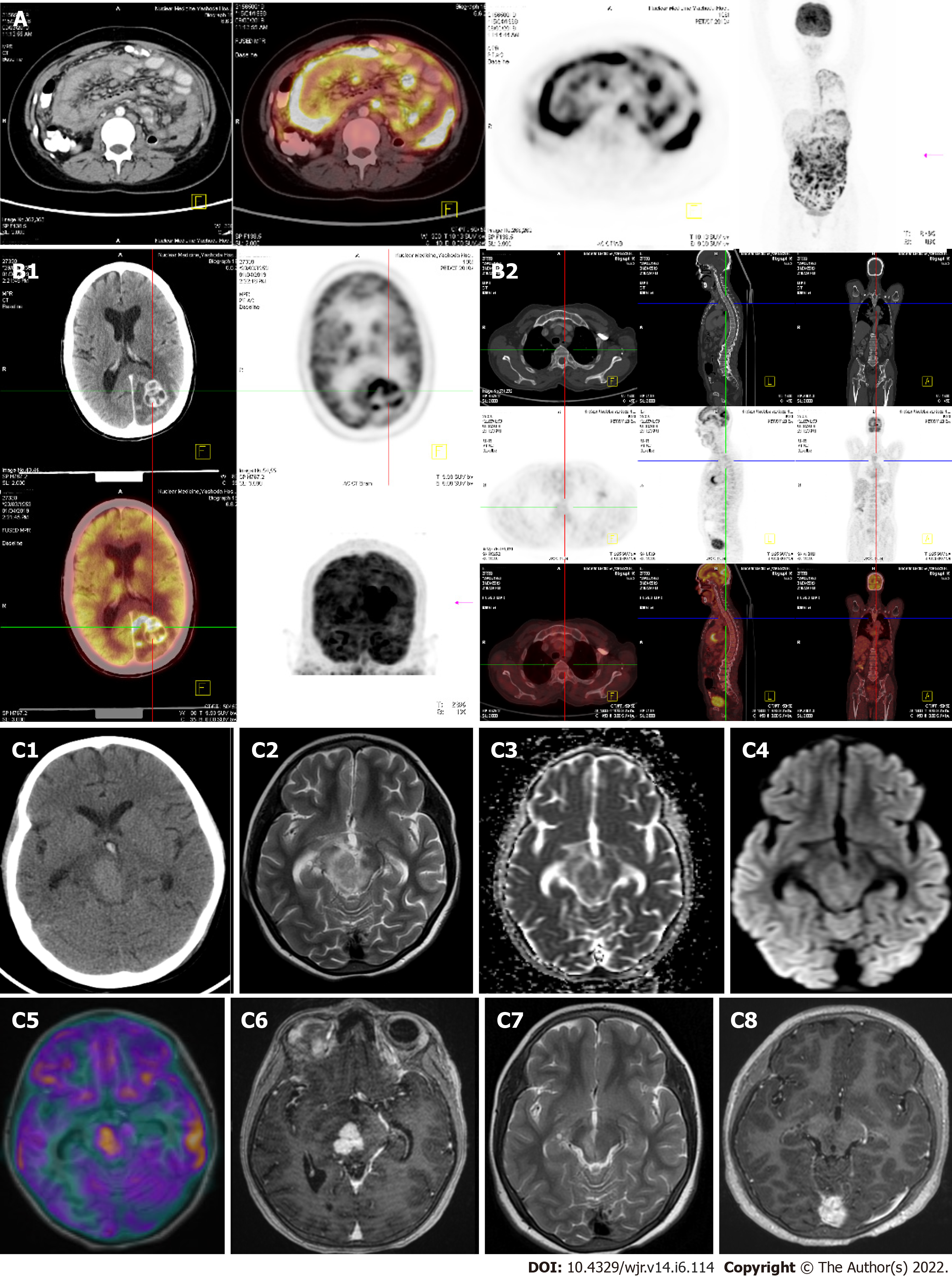
Figure 12 Fusion imaging.
A: Fluoro-deoxyglucose (FDG) positron emission tomography (PET)/computed tomography (CT) Abdominal tuberculosis: 55-year F - h/o loss of weight with mild abdominal pain on and off gradually increasing (for 4 mo). Low grade evening rise of fever. Whole body PET CT showing irregular peritoneal thickening with nodularities and cocoon formation. PET and fused PET-CT images showing significant amount of uptake with SUV max of 12.3; B: Whole body FDG-PET/CT - Brain Tuberculomas:45 years male - h/o seizures for 5 mo, gradually increasing in frequency. PET-CT advised for the possibility of metastases; B1: Whole body FDG-PET/CT done showing irregular ring enhancing lesions in the brain with peri-lesional edema. PET and fused PET-CT images showing significant amount of uptake with SUV max of 14.8; B2: There is no other abnormal uptake in the entire body. (Normal myocardial uptake and left axillary vessel uptake is noted); C: Fusion imaging (MR-PET): Tuberculoma with Rubral tremor. 12-year-old girl presented with right 3rd and 4th cranial nerve palsy along with rhythmic to and fro left ‘shoulder joint tremor’ which worsened with movement; C1: Axial non-contrast CT image demonstrates a well circumscribed hyperdense mass lesion within the right half of the midbrain; C2: T2-weighted imaging shows variable T2 hypo intensity within the lesion; C3 and C4: Diffusion weighted imaging and apparent diffusion co-efficient maps reveal restricted diffusion within the lesion; C5: Fusion imaging (T1W and PET) demonstrates avid glucose uptake within the lesion; C6: Post contrast T1 weighted imaging with fat saturation, reveals intense nodular enhancement. Stereotactic biopsy of the lesion revealed granulomatous inflammatory pathology; C7 and C8: After completion of anti-tuberculosis treatment, resolution of the granulomatous lesion with residual gliosis was observed on T2 weighted and Post contrast fat saturated T1w images. Images (A & B) courtesy Dr. Sikander Shaikh, Consultant radiologist, Yashodha Hospital, Hyderabad & Image (C) courtesy Dr. Saini J, Professor, Neuroimaging & IVR, NIMHANS, Bangalore.
- Citation: Merchant SA, Shaikh MJS, Nadkarni P. Tuberculosis conundrum - current and future scenarios: A proposed comprehensive approach combining laboratory, imaging, and computing advances. World J Radiol 2022; 14(6): 114-136
- URL: https://www.wjgnet.com/1949-8470/full/v14/i6/114.htm
- DOI: https://dx.doi.org/10.4329/wjr.v14.i6.114


