Copyright
©The Author(s) 2022.
World J Radiol. Jun 28, 2022; 14(6): 114-136
Published online Jun 28, 2022. doi: 10.4329/wjr.v14.i6.114
Published online Jun 28, 2022. doi: 10.4329/wjr.v14.i6.114
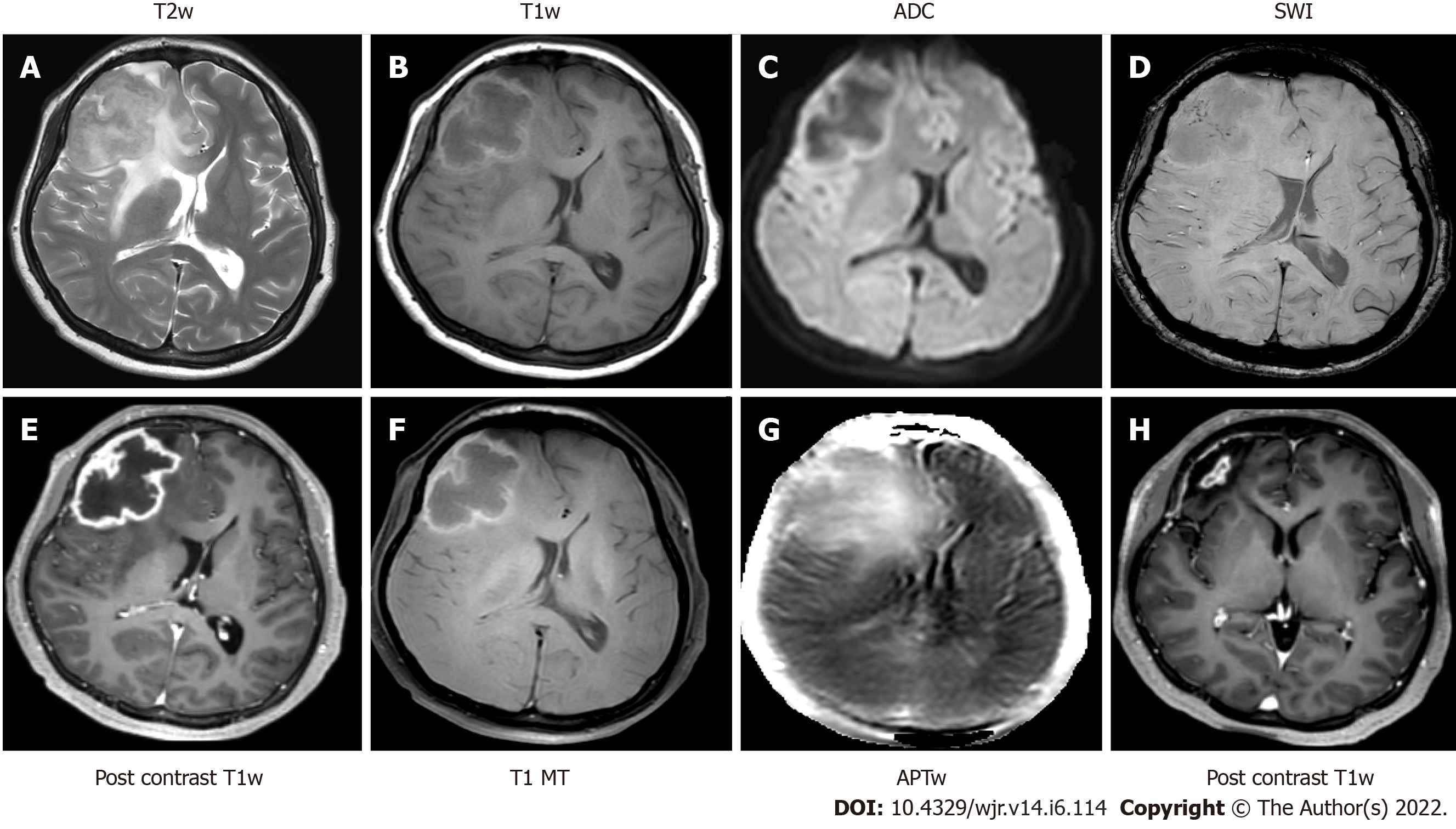
Figure 10 Magnetic resonance imaging - tuberculoma.
A: Axial T2-weighted imaging shows a variable T2 hypointense circumscribed mass lesion in the right anterior frontal region, with surrounding perilesional edema; B: T1 weighted imaging shows a peripheral T1 hyperintense rim; C: Apparent diffusion co-efficient map shows restriction of diffusion; D: Susceptibility weighted imaging demonstrates fine punctate intralesional foci of blooming; E Post contrast T1 weighted imaging showing slightly irregular peripheral rim enhancement; F: T1 magnetization transfer images; G: Amide proton transfer weighted images show elevated magnetization transfer asymmetry in the periphery of the lesion; H: T1 post contrast imaging after completion of anti-tuberculosis treatment reveals significant reduction in the size of the previously seen ring enhancing lesion. APT: Amide proton transfer. Images courtesy Dr. Saini J, Professor, Neuroimaging & IVR, NIMHANS, Bangalore.
- Citation: Merchant SA, Shaikh MJS, Nadkarni P. Tuberculosis conundrum - current and future scenarios: A proposed comprehensive approach combining laboratory, imaging, and computing advances. World J Radiol 2022; 14(6): 114-136
- URL: https://www.wjgnet.com/1949-8470/full/v14/i6/114.htm
- DOI: https://dx.doi.org/10.4329/wjr.v14.i6.114


