Copyright
©The Author(s) 2022.
World J Radiol. Jun 28, 2022; 14(6): 114-136
Published online Jun 28, 2022. doi: 10.4329/wjr.v14.i6.114
Published online Jun 28, 2022. doi: 10.4329/wjr.v14.i6.114
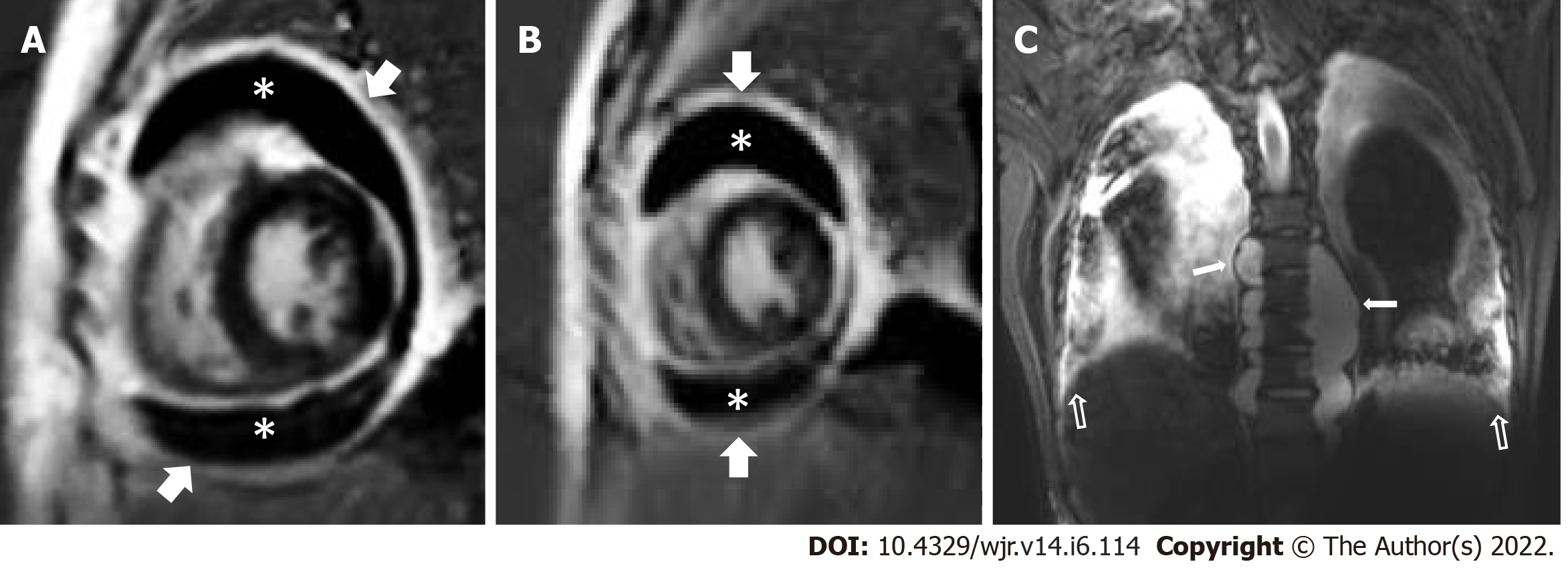
Figure 8 Tuberculous pericarditis and pericardial effusion: 3 Tesla Cardiac magnetic resonance imaging.
A and B: PSIR (short axis view) images shows enhancing pericardial thickening (arrow) and moderate distension of pericardial space with hypointense fluid (asterisk); C: Coronal STIR dorsal spine: Paraspinal abscesses (white arrow -filled) with concomitant pleural effusions (white arrow- unfilled).
- Citation: Merchant SA, Shaikh MJS, Nadkarni P. Tuberculosis conundrum - current and future scenarios: A proposed comprehensive approach combining laboratory, imaging, and computing advances. World J Radiol 2022; 14(6): 114-136
- URL: https://www.wjgnet.com/1949-8470/full/v14/i6/114.htm
- DOI: https://dx.doi.org/10.4329/wjr.v14.i6.114


