Copyright
©The Author(s) 2022.
World J Radiol. Jun 28, 2022; 14(6): 114-136
Published online Jun 28, 2022. doi: 10.4329/wjr.v14.i6.114
Published online Jun 28, 2022. doi: 10.4329/wjr.v14.i6.114
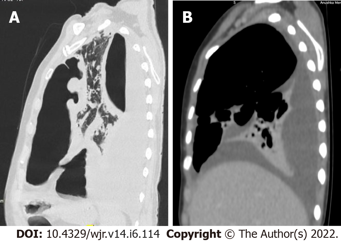
Figure 5 Tuberculous pyo-pneumothorax.
A: Sagittal high-resolution computed tomography image in lung window showing a thick-walled cavity communicating with the left pleural space. A large loculated collection in the left pleural space showing air-fluid level; B: Sagittal image in a mediastinal window showing a Right pleural effusion with partial collapse of Right lower lobe. Images courtesy Dr. Thakkar H, Prof & Head (Radiology), KEM Hospital, Mumbai.
- Citation: Merchant SA, Shaikh MJS, Nadkarni P. Tuberculosis conundrum - current and future scenarios: A proposed comprehensive approach combining laboratory, imaging, and computing advances. World J Radiol 2022; 14(6): 114-136
- URL: https://www.wjgnet.com/1949-8470/full/v14/i6/114.htm
- DOI: https://dx.doi.org/10.4329/wjr.v14.i6.114


