Copyright
©The Author(s) 2022.
World J Radiol. Jun 28, 2022; 14(6): 114-136
Published online Jun 28, 2022. doi: 10.4329/wjr.v14.i6.114
Published online Jun 28, 2022. doi: 10.4329/wjr.v14.i6.114
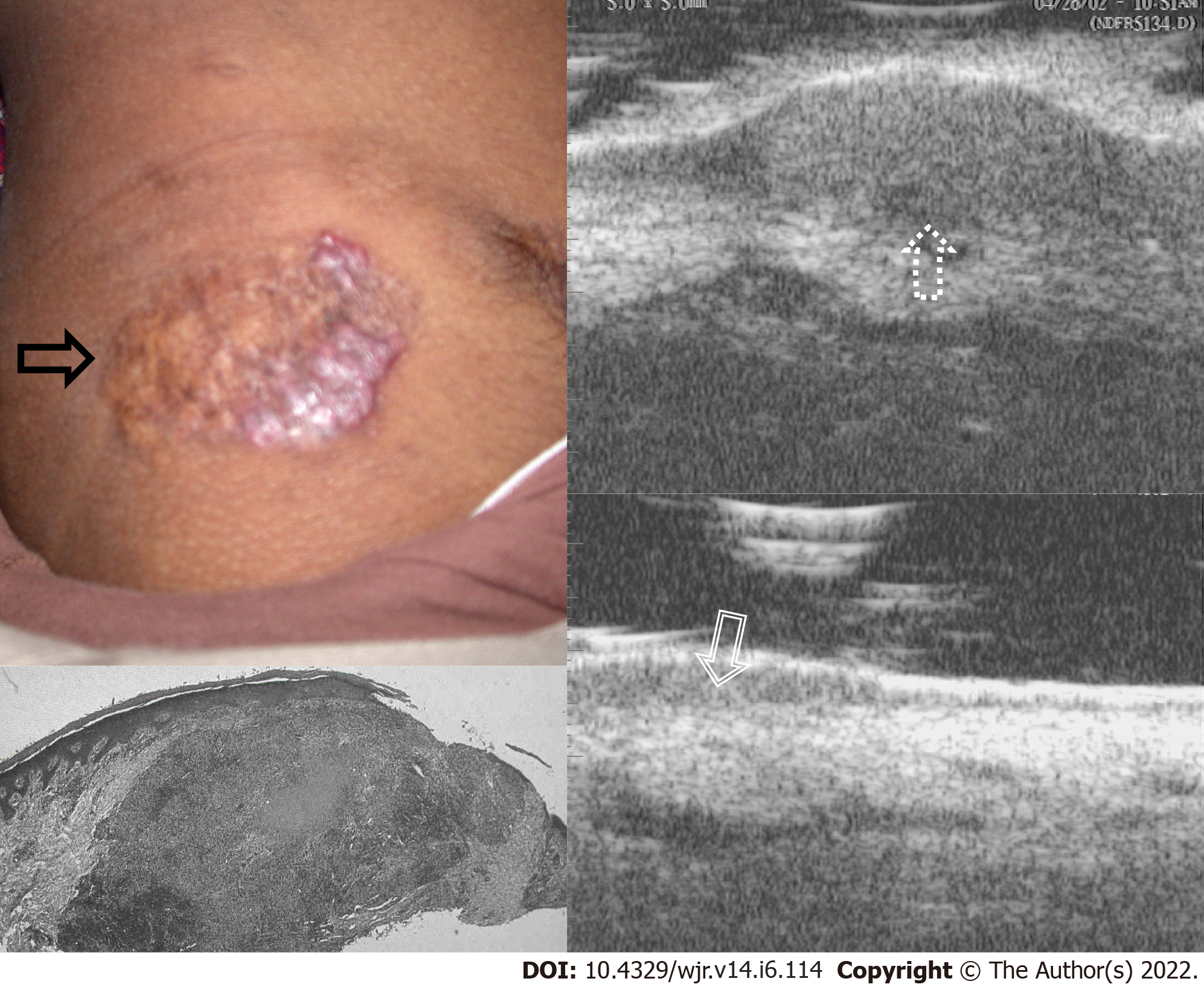
Figure 4 Ultrasound Biomicroscopy scanned at 50 MHz - Skin tuberculosis - lupus vulgaris.
A well-defined reddish-brown plaque with papulo-nodular borders is seen on the skin (black arrow), Ultrasound biomicroscopy (UBM) shows a well-defined heterogenous mass lesion in the dermis (up arrow-dotted), Histopathology shows a well-defined tuberculous granuloma in the dermis (white filled arrow), Follow up UBM after 6 mo of AKT shows marked decrease in the size of granuloma in the dermis (down arrow- dashed). Images Courtesy Dr. Bhatt K, UBM Institute & Sonography Centre, Mumbai.
- Citation: Merchant SA, Shaikh MJS, Nadkarni P. Tuberculosis conundrum - current and future scenarios: A proposed comprehensive approach combining laboratory, imaging, and computing advances. World J Radiol 2022; 14(6): 114-136
- URL: https://www.wjgnet.com/1949-8470/full/v14/i6/114.htm
- DOI: https://dx.doi.org/10.4329/wjr.v14.i6.114


