Copyright
©The Author(s) 2022.
World J Radiol. Jun 28, 2022; 14(6): 114-136
Published online Jun 28, 2022. doi: 10.4329/wjr.v14.i6.114
Published online Jun 28, 2022. doi: 10.4329/wjr.v14.i6.114
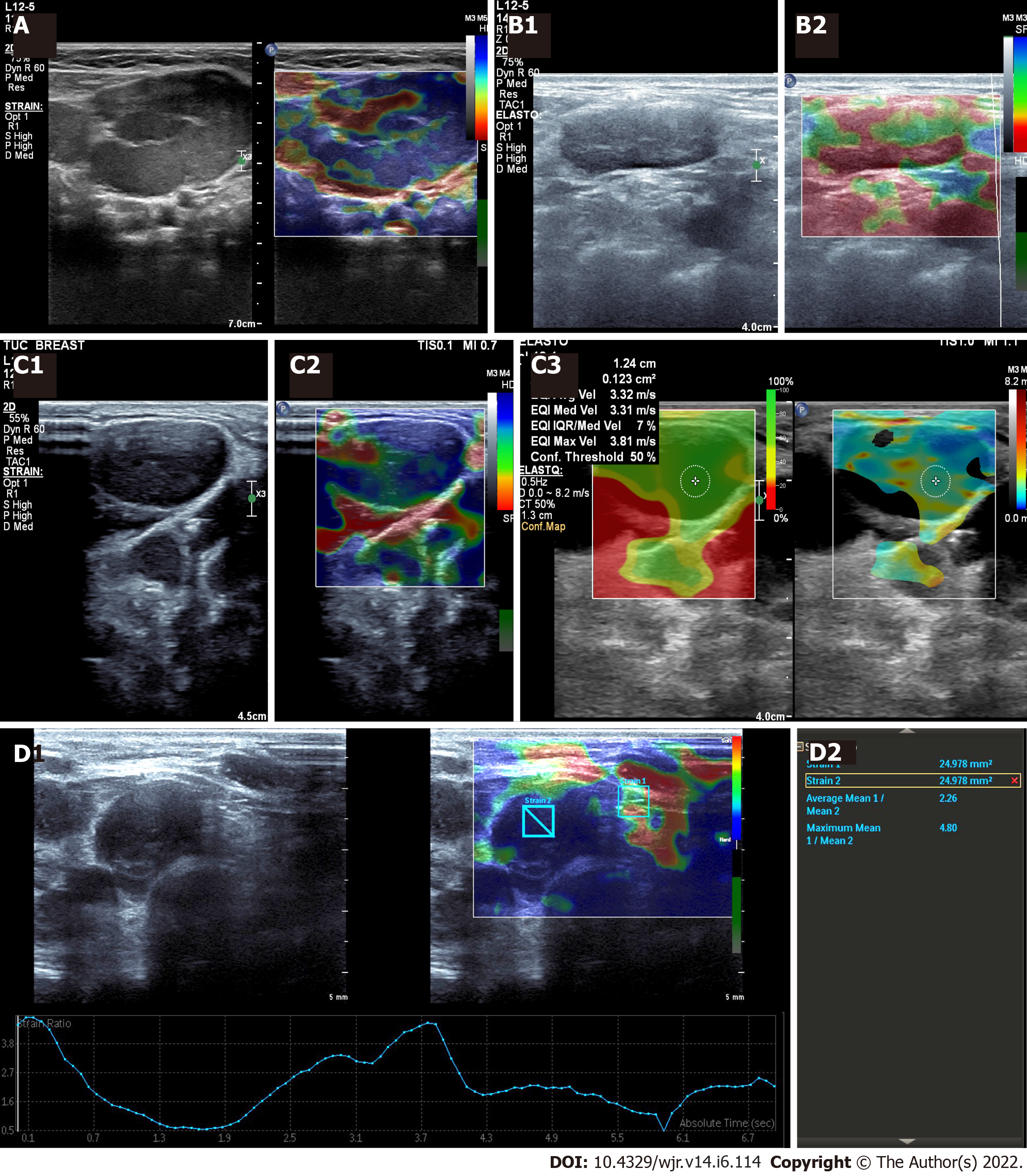
Figure 2 Cervical tuberculosis lymph node.
A: Cervical tuberculosis lymph node: Ultrasonography (US) elastography - central necrotic area appears soft (red); B: Tuberculous lymphadenopathy: A 16-year-old female with fever and neck swelling; B1: Grey scale B-mode image: shows an enlarged lymph node with diffusely hypoechoic echotexture and loss of fatty hilum; B2: Strain US Elastography: Showing a mixed pattern, predominantly soft (red); C: Tuberculous lymphadenopathy: 35-year-old male with neck swelling and history of weight loss; C1: Grey scale image: shows an enlarged lymph node with diffusely hypoechoic echotexture and loss of fatty hilum; C2: Strain US elastography: Showing soft areas within (red areas) s/o necrosis / liquefaction; C3: Shear wave US elastography: Shows relatively low shear wave values; D: Tuberculous lymphadenopathy: Neck US of a 14-year-old female (known case of drug resistant tuberculosis); D1: Grey scale B-mode image: enlarged lymph nodes with diffusely hypoechoic echotexture and loss of fatty hilum; D2: Strain US Elastography: The strain elastography reveals a low strain ratio (2.26). Elastography details are noted on the elastography graph too. Trucut biopsy was done - results awaited. Images courtesy Dr. Chaubal N, Thane Ultrasound Centre, India.
- Citation: Merchant SA, Shaikh MJS, Nadkarni P. Tuberculosis conundrum - current and future scenarios: A proposed comprehensive approach combining laboratory, imaging, and computing advances. World J Radiol 2022; 14(6): 114-136
- URL: https://www.wjgnet.com/1949-8470/full/v14/i6/114.htm
- DOI: https://dx.doi.org/10.4329/wjr.v14.i6.114


