Copyright
©The Author(s) 2022.
World J Radiol. Jun 28, 2022; 14(6): 114-136
Published online Jun 28, 2022. doi: 10.4329/wjr.v14.i6.114
Published online Jun 28, 2022. doi: 10.4329/wjr.v14.i6.114
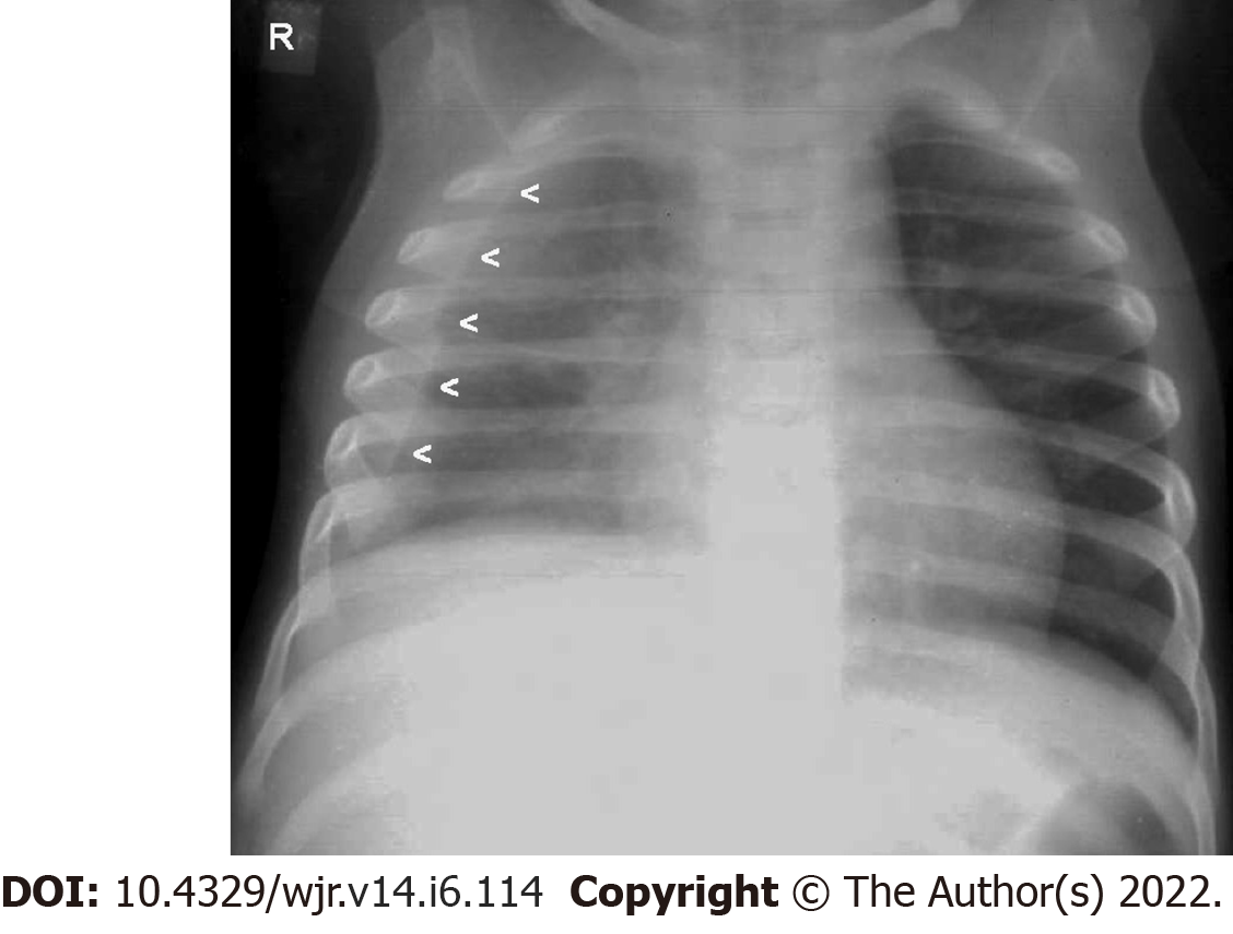
Figure 1 Lamellar pleural effusion.
Frontal chest radiograph of an 18-mo-old child with Pulmonary tuberculosis (primary complex) reveals a lamellar pleural effusion- (homogeneous increased radio-opacity along lateral aspect of right lung field with blunting of the right costophrenic angle- mimicking the appearance of pleural thickening) - [arrowheads]. Image courtesy – Department of Radiology, KEM Hospital, Mumbai.
- Citation: Merchant SA, Shaikh MJS, Nadkarni P. Tuberculosis conundrum - current and future scenarios: A proposed comprehensive approach combining laboratory, imaging, and computing advances. World J Radiol 2022; 14(6): 114-136
- URL: https://www.wjgnet.com/1949-8470/full/v14/i6/114.htm
- DOI: https://dx.doi.org/10.4329/wjr.v14.i6.114


