Copyright
©The Author(s) 2022.
World J Radiol. Apr 28, 2022; 14(4): 91-103
Published online Apr 28, 2022. doi: 10.4329/wjr.v14.i4.91
Published online Apr 28, 2022. doi: 10.4329/wjr.v14.i4.91
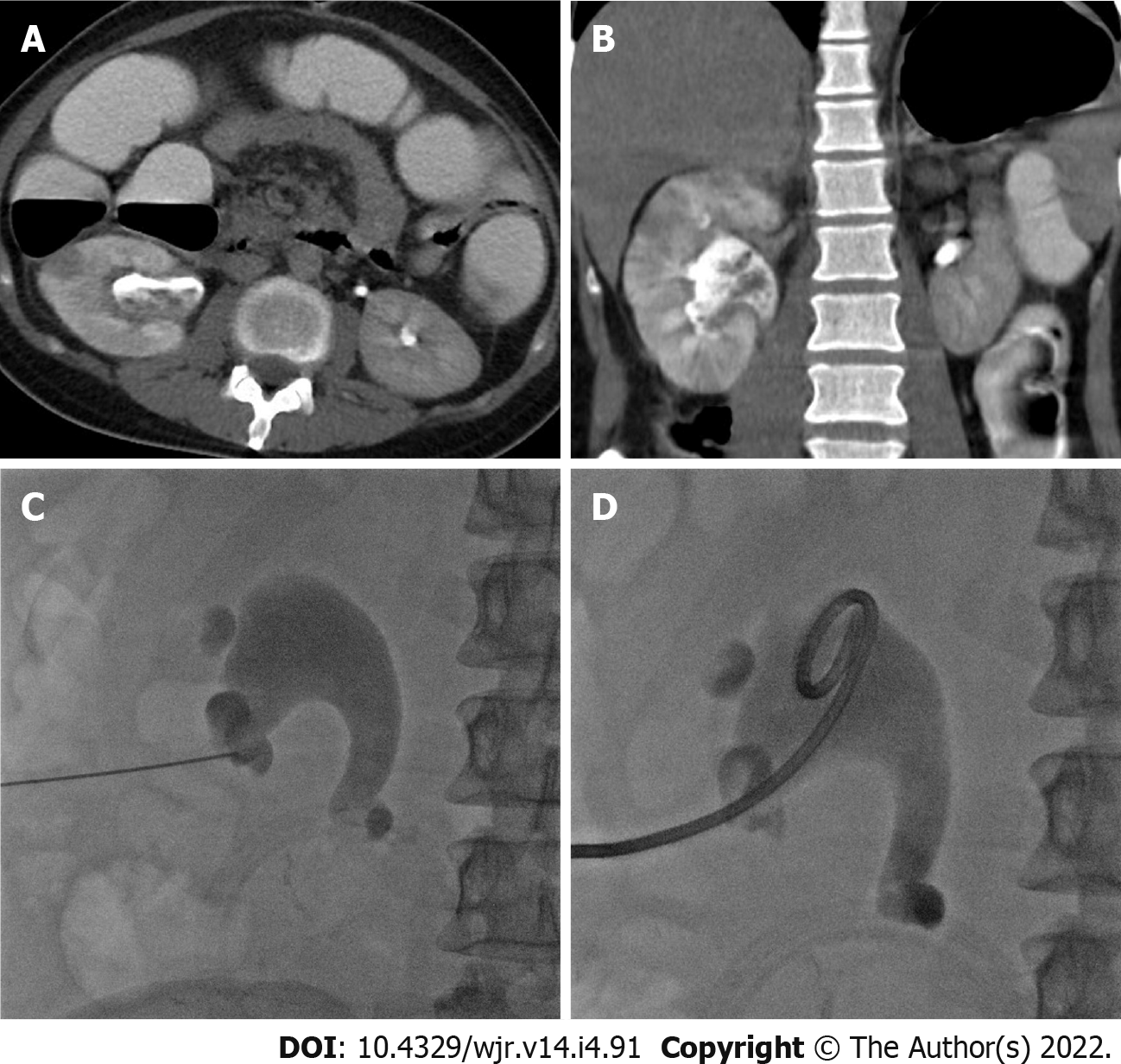
Figure 6 Percutaneous nephrostomy in a 30-yr-old male presented with acute pyelonephritis.
A and B: Axial and coronal computed tomography images in excretory phase show characteristic features of acute pyelonephritis in the form of focal hypoenhnacing areas (striated nephrogram) and debris in dilated renal pelvis; C and D: Frontal fluoroscopic images show puncture needle in the lower calyces and successful insertion of nephrostomy tube.
- Citation: Deif MA, Mounir AM, Abo-Hedibah SA, Abdel Khalek AM, Elmokadem AH. Outcome of percutaneous drainage for septic complications coexisted with COVID-19. World J Radiol 2022; 14(4): 91-103
- URL: https://www.wjgnet.com/1949-8470/full/v14/i4/91.htm
- DOI: https://dx.doi.org/10.4329/wjr.v14.i4.91


