Copyright
©The Author(s) 2022.
World J Radiol. Apr 28, 2022; 14(4): 91-103
Published online Apr 28, 2022. doi: 10.4329/wjr.v14.i4.91
Published online Apr 28, 2022. doi: 10.4329/wjr.v14.i4.91
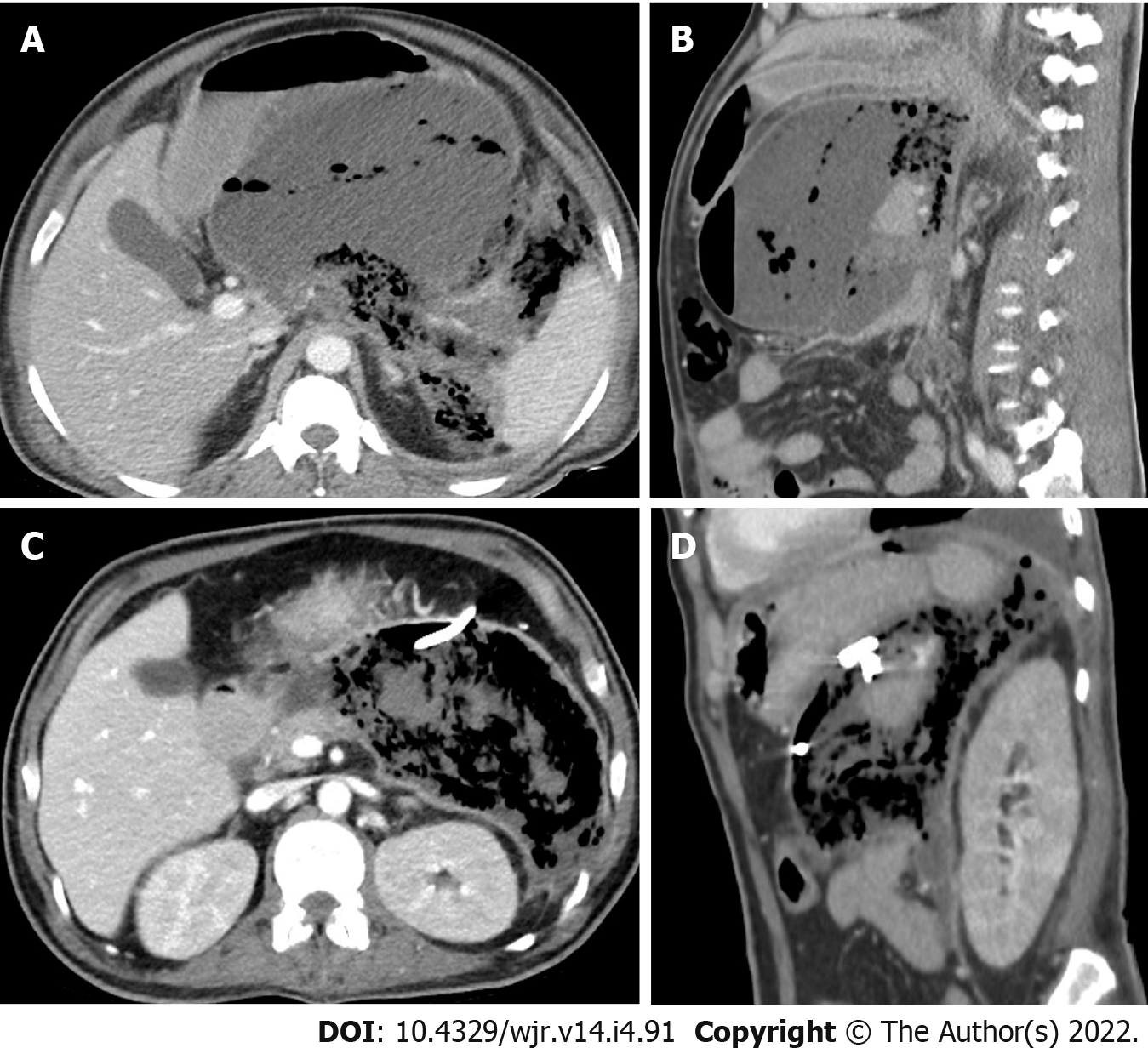
Figure 3 Percutaneous drainage of peripancreatic collection in a 43-yr-old male presented by acute pancreatitis.
A and B: Axial and sagittal contrast enhanced computed tomography (CT) images show large peripancreatic collection/walled-off necrosis. The collection is mixed with pockets of gas inside and there is extension of the gas density into the retroperitoneal and perisplenic spaces; C and D: Axial and sagittal contrast enhanced CT images 22 d after tube insertion show reduction of the collection size with increased amount of gas within the collection.
- Citation: Deif MA, Mounir AM, Abo-Hedibah SA, Abdel Khalek AM, Elmokadem AH. Outcome of percutaneous drainage for septic complications coexisted with COVID-19. World J Radiol 2022; 14(4): 91-103
- URL: https://www.wjgnet.com/1949-8470/full/v14/i4/91.htm
- DOI: https://dx.doi.org/10.4329/wjr.v14.i4.91


