Copyright
©The Author(s) 2022.
World J Radiol. Apr 28, 2022; 14(4): 91-103
Published online Apr 28, 2022. doi: 10.4329/wjr.v14.i4.91
Published online Apr 28, 2022. doi: 10.4329/wjr.v14.i4.91
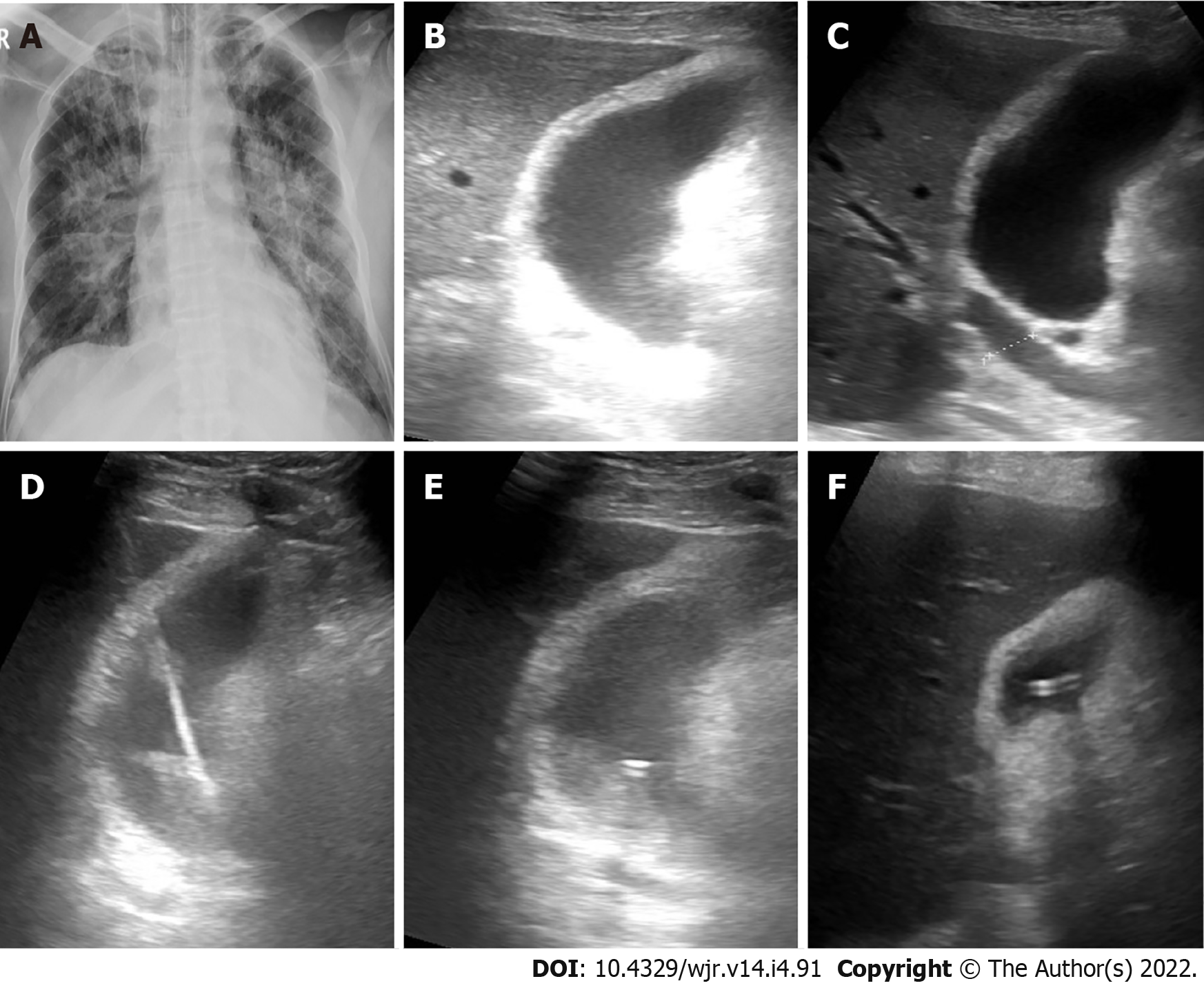
Figure 2 Cholecystostomy in a 72-yr-old male presented by acute cholecystitis.
A: Frontal chest X-ray shows opacities involving both lungs with central predominance; B and C: B-mode ultrasound images show distended thick-walled gall bladder with biliary dilatation; D: B-mode ultrasound image show puncture needle through the gall bladder; E: B-mode ultrasound image tube inside the gall bladder; F: B-mode ultrasound image of the gall bladder after drainage.
- Citation: Deif MA, Mounir AM, Abo-Hedibah SA, Abdel Khalek AM, Elmokadem AH. Outcome of percutaneous drainage for septic complications coexisted with COVID-19. World J Radiol 2022; 14(4): 91-103
- URL: https://www.wjgnet.com/1949-8470/full/v14/i4/91.htm
- DOI: https://dx.doi.org/10.4329/wjr.v14.i4.91


