Copyright
©The Author(s) 2022.
World J Radiol. Apr 28, 2022; 14(4): 70-81
Published online Apr 28, 2022. doi: 10.4329/wjr.v14.i4.70
Published online Apr 28, 2022. doi: 10.4329/wjr.v14.i4.70
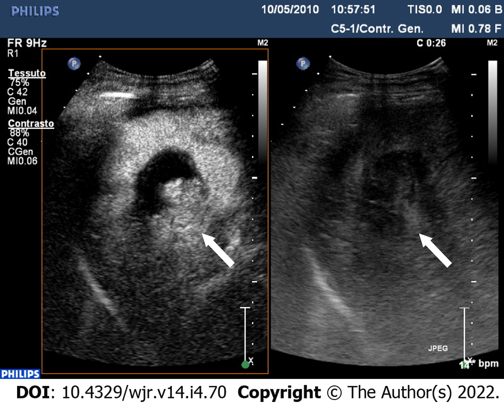
Figure 4 One-month post-procedural assessment of hepatocellular carcinoma after TACE.
Contrast-enhanced ultrasound examination in the arterial phase (26 s after the i.v. injection of contrast agent) shows a clear cut intralesional area of contrast-enhancement indicating still viable tumor.
- Citation: Bartolotta TV, Randazzo A, Bruno E, Taibbi A. Focal liver lesions in cirrhosis: Role of contrast-enhanced ultrasonography. World J Radiol 2022; 14(4): 70-81
- URL: https://www.wjgnet.com/1949-8470/full/v14/i4/70.htm
- DOI: https://dx.doi.org/10.4329/wjr.v14.i4.70


