Copyright
©The Author(s) 2022.
World J Radiol. Feb 28, 2022; 14(2): 30-46
Published online Feb 28, 2022. doi: 10.4329/wjr.v14.i2.30
Published online Feb 28, 2022. doi: 10.4329/wjr.v14.i2.30
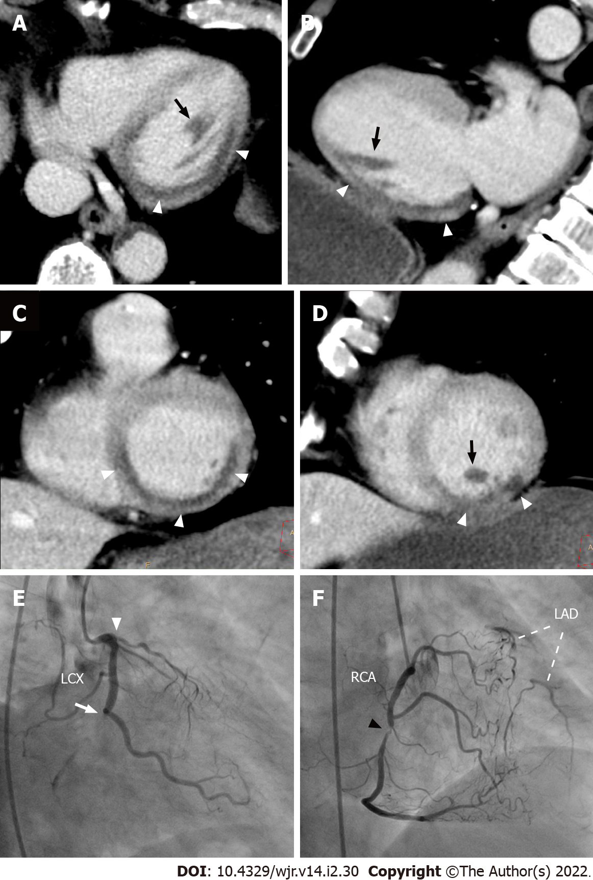
Figure 16 Myocardial perfusion defect of the posteromedial papillary muscle on non-electrocardiogram-gated contrast-enhanced computed tomography.
A 68-year-old man with right anterior chest pain underwent non-electrocardiogram (ECG)-gated contrast-enhanced computed tomography (CECT) in search of aortic dissection. Axial (A), vertical long axis (B), and short axis (C, D) reformatted non-ECG-gated CECT images acquired 120 s after contrast injection showed decreased myocardial enhancement in the basal to mid inferior, inferolateral, and inferoseptal walls of the left ventricle (arrowheads). Decreased myocardial enhancement was also recognized in the posteromedial papillary muscle (arrows). Invasive coronary angiography showed total occlusion in the distal site of the left circumflex coronary artery (E, arrow), total occlusion in the proximal site of the left anterior descending artery (E, arrowhead), and 90% stenosis in the mid site of the right coronary artery (F, arrowhead), which provides abundant collateral flow to the left anterior descending artery. The patient subsequently underwent an emergent coronary artery bypass grafting. LAD: Left anterior descending artery; LCX: Left circumflex coronary artery; RCA: Right coronary artery.
- Citation: Yoshihara S. Acute coronary syndrome on non-electrocardiogram-gated contrast-enhanced computed tomography. World J Radiol 2022; 14(2): 30-46
- URL: https://www.wjgnet.com/1949-8470/full/v14/i2/30.htm
- DOI: https://dx.doi.org/10.4329/wjr.v14.i2.30


