Copyright
©The Author(s) 2022.
World J Radiol. Feb 28, 2022; 14(2): 30-46
Published online Feb 28, 2022. doi: 10.4329/wjr.v14.i2.30
Published online Feb 28, 2022. doi: 10.4329/wjr.v14.i2.30
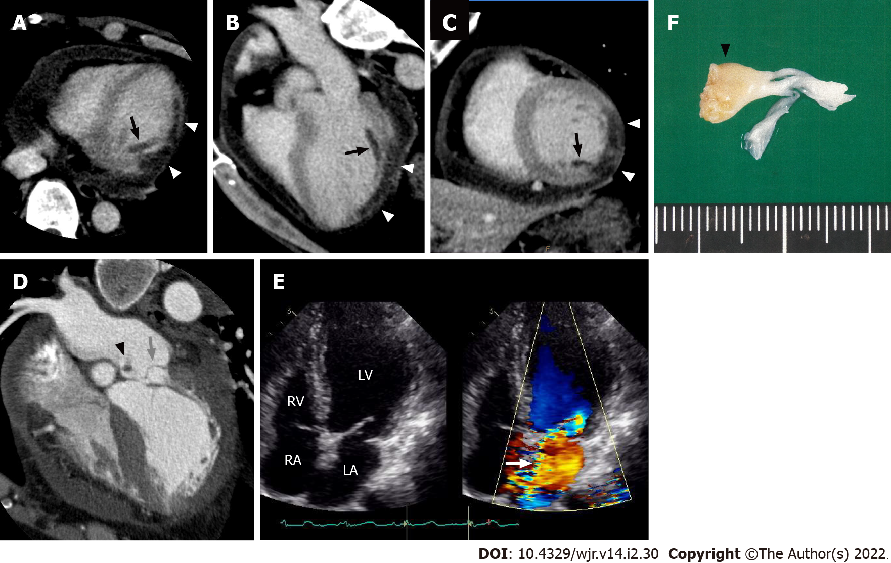
Figure 15 Postinfarct left ventricular papillary muscle rupture.
Electrocardiogram-gated cardiac CT images from a 61-year-old man with posteromedial papillary muscle rupture complicated by acute ST elevation myocardial infarction due to total occlusion of the left circumflex coronary artery. Axial (A), three-chamber (B), and short axis (C) reformatted cardiac CT images showed decreased myocardial enhancement in the inferolateral wall (white arrowheads) and posteromedial papillary muscle (black arrows) of the left ventricle. A horizontal long axis image (D) showed severe prolapse of the posterior mitral valve leaflet into the left atrium (grey arrow) with discernible papillary muscle attachment (black arrowhead). An apical four-chamber color Doppler image of the transthoracic echocardiogram showed severe mitral regurgitation during ventricular systole extending to the posterior left atrial wall (E, white arrow). The patient subsequently underwent a mitral valve replacement. Surgical specimen showed posteromedial papillary muscle attached to the resected posterior mitral leaflet (F, black arrowhead). LV: Left ventricle; LA: Left atrium; RV: Right ventricle; RA: Right atrium.
- Citation: Yoshihara S. Acute coronary syndrome on non-electrocardiogram-gated contrast-enhanced computed tomography. World J Radiol 2022; 14(2): 30-46
- URL: https://www.wjgnet.com/1949-8470/full/v14/i2/30.htm
- DOI: https://dx.doi.org/10.4329/wjr.v14.i2.30


