Copyright
©The Author(s) 2022.
World J Radiol. Feb 28, 2022; 14(2): 30-46
Published online Feb 28, 2022. doi: 10.4329/wjr.v14.i2.30
Published online Feb 28, 2022. doi: 10.4329/wjr.v14.i2.30
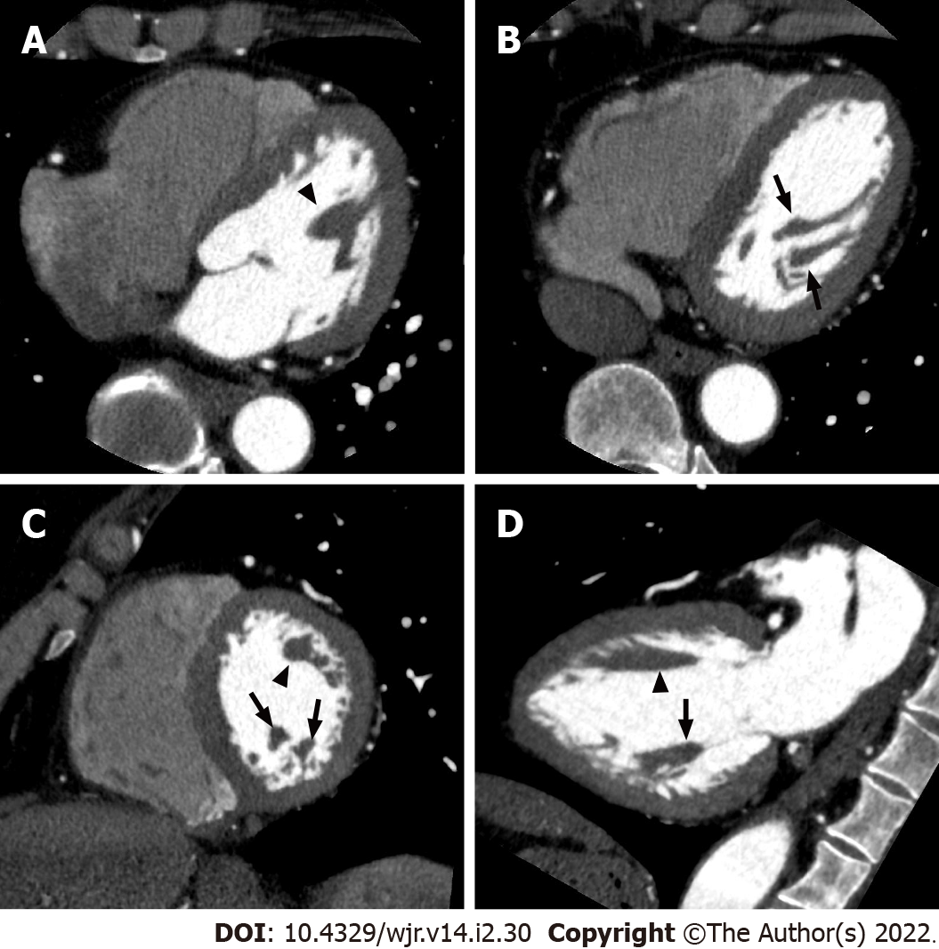
Figure 14 Left ventricular papillary muscles.
Axial (A, B), short axis (C), and vertical long axis (D) reformatted electrocardiogram-gated cardiac CT images showing the anatomy of the left ventricular chamber of the normal heart. The anterolateral (arrowheads) and posteromedial (arrows) papillary muscles manifest as filling defects within the contrast-filled left ventricular lumen.
- Citation: Yoshihara S. Acute coronary syndrome on non-electrocardiogram-gated contrast-enhanced computed tomography. World J Radiol 2022; 14(2): 30-46
- URL: https://www.wjgnet.com/1949-8470/full/v14/i2/30.htm
- DOI: https://dx.doi.org/10.4329/wjr.v14.i2.30


