Copyright
©The Author(s) 2022.
World J Radiol. Feb 28, 2022; 14(2): 30-46
Published online Feb 28, 2022. doi: 10.4329/wjr.v14.i2.30
Published online Feb 28, 2022. doi: 10.4329/wjr.v14.i2.30
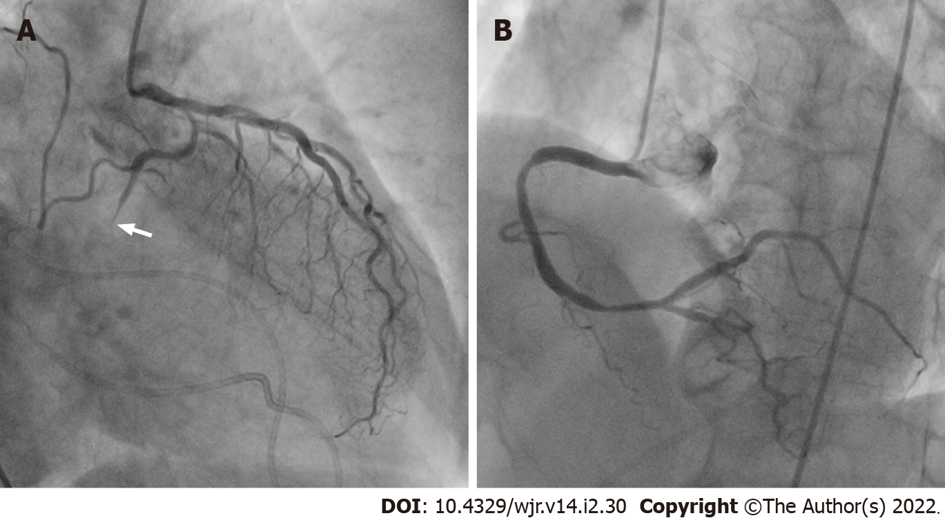
Figure 13 Corresponding invasive coronary angiography in postinfarct cardiac free wall rupture.
Same patient as Figure 12. A: Invasive coronary angiography showed total occlusion in the distal site of the left circumflex coronary artery (arrow); B: The right coronary artery showed no significant stenosis. The patient subsequently underwent a surgical operation. After removal of the large clot within the pericardium, a small perforation was found in the lateral wall of the left ventricle, confirming a definitive diagnosis of left ventricular free wall rupture.
- Citation: Yoshihara S. Acute coronary syndrome on non-electrocardiogram-gated contrast-enhanced computed tomography. World J Radiol 2022; 14(2): 30-46
- URL: https://www.wjgnet.com/1949-8470/full/v14/i2/30.htm
- DOI: https://dx.doi.org/10.4329/wjr.v14.i2.30


