Copyright
©The Author(s) 2022.
World J Radiol. Feb 28, 2022; 14(2): 30-46
Published online Feb 28, 2022. doi: 10.4329/wjr.v14.i2.30
Published online Feb 28, 2022. doi: 10.4329/wjr.v14.i2.30
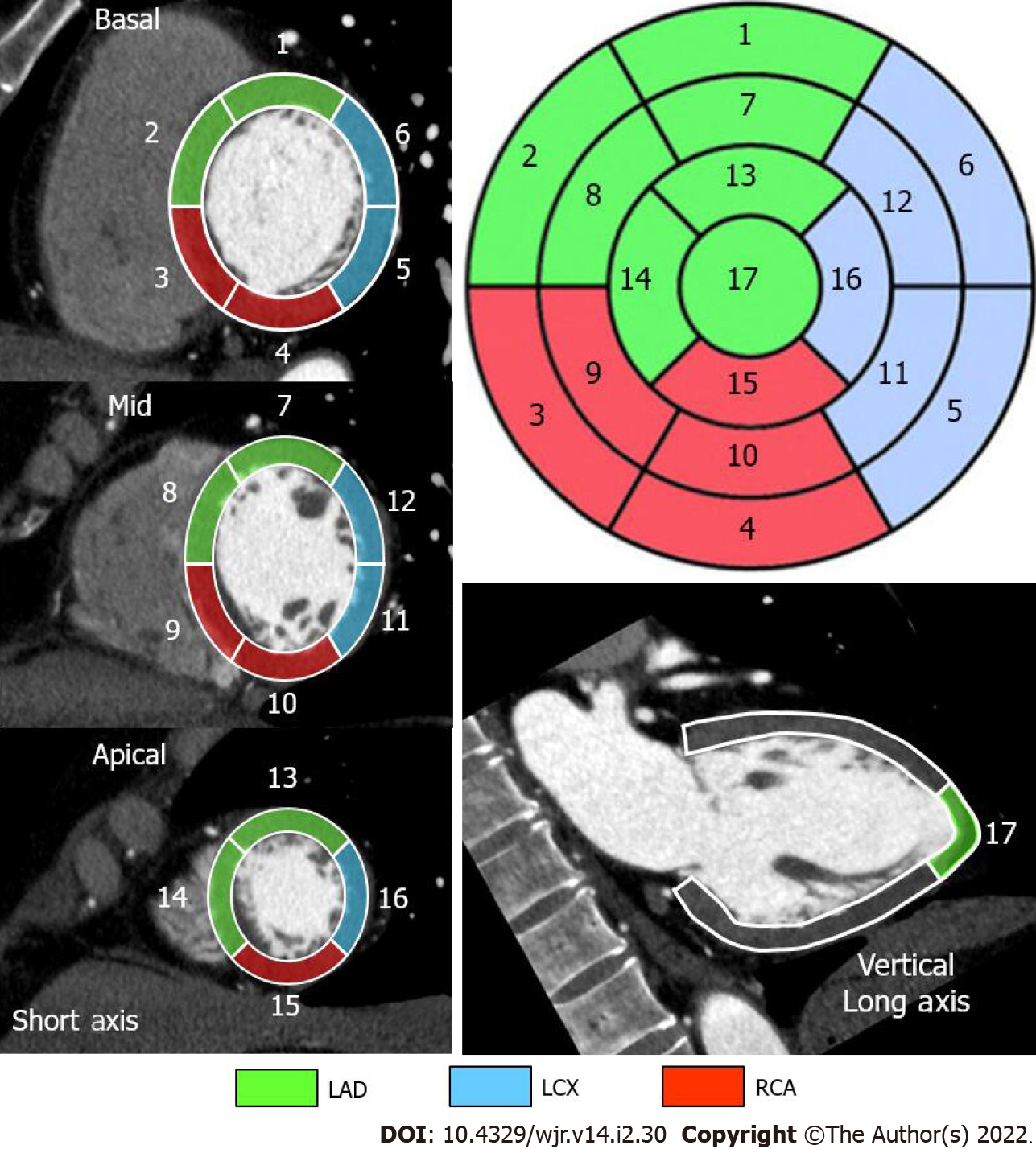
Figure 2 Standard segmental myocardial display in a 17-segment model.
Electrocardiogram-gated cardiac computed tomography-based individual assignment of left ventricular segmentation following the American Heart Association 17-segment model with corresponding color-coded coronary artery perfusion territories for a right-dominant coronary system. LAD: Left anterior descending artery in green; LCX: Left circumflex coronary artery in blue; RCA: Right coronary artery in red.
- Citation: Yoshihara S. Acute coronary syndrome on non-electrocardiogram-gated contrast-enhanced computed tomography. World J Radiol 2022; 14(2): 30-46
- URL: https://www.wjgnet.com/1949-8470/full/v14/i2/30.htm
- DOI: https://dx.doi.org/10.4329/wjr.v14.i2.30


