Copyright
©The Author(s) 2022.
World J Radiol. Feb 28, 2022; 14(2): 30-46
Published online Feb 28, 2022. doi: 10.4329/wjr.v14.i2.30
Published online Feb 28, 2022. doi: 10.4329/wjr.v14.i2.30
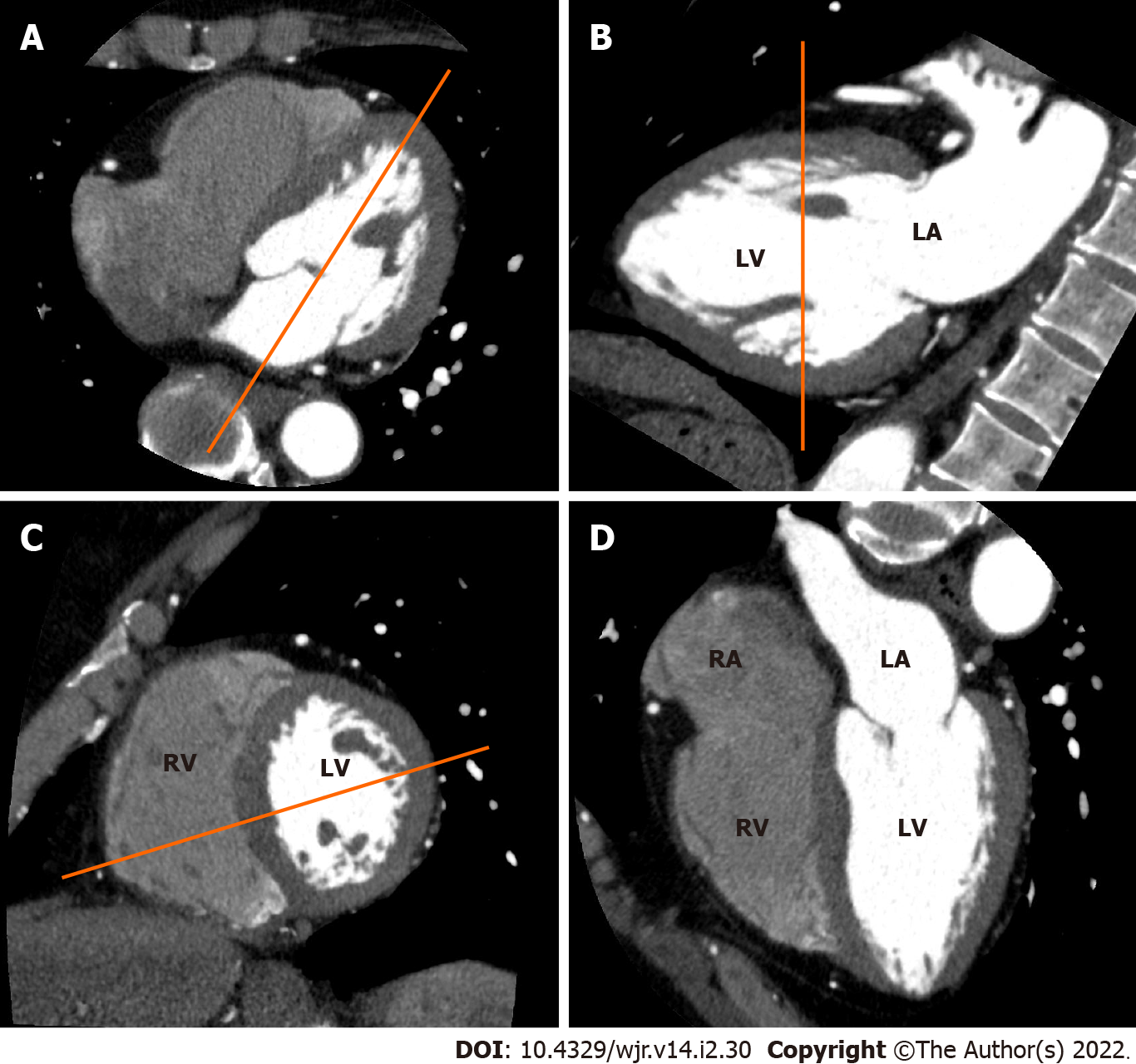
Figure 1 Electrocardiogram-gated cardiac computed tomography (CT)-based sequential approach to CT imaging of cardiac planes.
A: From an axial CT data set at the level of the mitral valve, a longitudinal plane bisecting the mitral valve and the left ventricular apex is used to create a vertical long axis view; B: From the vertical long axis view, a slice parallel to the mitral annulus at the mid ventricular level is used to obtain a short axis view; C: From the short axis view, a slice bisecting the center of the left ventricle (LV) and the intersection between the junction of the free wall and diaphragmatic wall of the right ventricle (RV) are used to obtain a horizontal long axis view. (D) Horizontal long axis view showing the left atrium, LV, right atrium and RV.
- Citation: Yoshihara S. Acute coronary syndrome on non-electrocardiogram-gated contrast-enhanced computed tomography. World J Radiol 2022; 14(2): 30-46
- URL: https://www.wjgnet.com/1949-8470/full/v14/i2/30.htm
- DOI: https://dx.doi.org/10.4329/wjr.v14.i2.30


