Copyright
©The Author(s) 2022.
World J Radiol. Nov 28, 2022; 14(11): 367-374
Published online Nov 28, 2022. doi: 10.4329/wjr.v14.i11.367
Published online Nov 28, 2022. doi: 10.4329/wjr.v14.i11.367
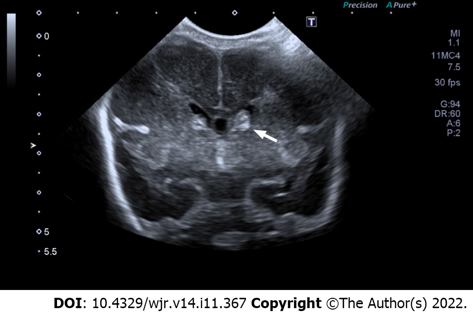
Figure 2 Grade II intraventricular hemorrhage.
Coronal view ultrasound image reveals abnormal echogenicity in the left caudothalamic groove extending into left lateral ventricle. There was no associated ventricular dilatation (white arrow).
- Citation: Barakzai MD, Khalid A, Sheer ZZ, Khan F, Nadeem N, Khan N, Hilal K. Interobserver reliability between pediatric radiologists and residents in ultrasound evaluation of intraventricular hemorrhage in premature infants. World J Radiol 2022; 14(11): 367-374
- URL: https://www.wjgnet.com/1949-8470/full/v14/i11/367.htm
- DOI: https://dx.doi.org/10.4329/wjr.v14.i11.367


