Copyright
©The Author(s) 2021.
World J Radiol. Sep 28, 2021; 13(9): 307-313
Published online Sep 28, 2021. doi: 10.4329/wjr.v13.i9.307
Published online Sep 28, 2021. doi: 10.4329/wjr.v13.i9.307
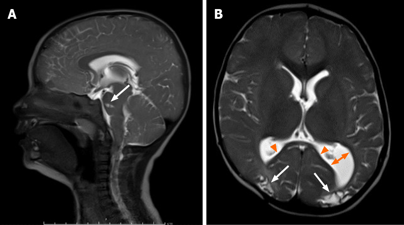
Figure 6 Follow-up magnetic resonance imaging at 15 mo.
A: Follow-up magnetic resonance imaging at 15 mo demonstrates continued resolution of the subdural hematomas and obstructive hydrocephalus on sagittal T2. Note the focal encephalomalacia at the pons (arrow); B: Axial T2 demonstrates encephalomalacic change manifested by thinning of the posterior corpus callosum (arrowheads), decreased gray and white matter of the posterior occipital regions bilaterally (arrows), and colpocephaly of the left lateral ventricle (two direction arrow).
- Citation: Rousslang LK, Rooks EA, Meldrum JT, Hooten KG, Wood JR. Neonatal infratentorial subdural hematoma contributing to obstructive hydrocephalus in the setting of therapeutic cooling: A case report. World J Radiol 2021; 13(9): 307-313
- URL: https://www.wjgnet.com/1949-8470/full/v13/i9/307.htm
- DOI: https://dx.doi.org/10.4329/wjr.v13.i9.307


