Copyright
©The Author(s) 2021.
World J Radiol. Sep 28, 2021; 13(9): 307-313
Published online Sep 28, 2021. doi: 10.4329/wjr.v13.i9.307
Published online Sep 28, 2021. doi: 10.4329/wjr.v13.i9.307
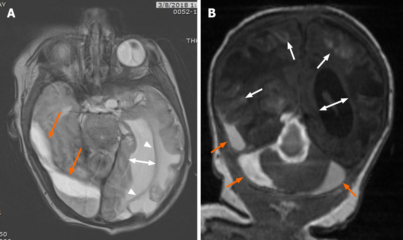
Figure 4 Follow-up magnetic resonance imaging on 20th day of life.
A: Follow-up magnetic resonance imaging on 20th day of life revealed evolving blood products (orange arrows) in the subdural space on axial T2, with interval left greater than right cystic encephalomalacia in the parietal and occipital lobes and left greater than right ex-vacuo dilatation of the lateral ventricles (two direction arrow); B: Coronal T1 demonstrates degrading blood product in the right temporal lobe subdural space, and central and peripheral infratentorial subdural spaces (orange arrows) with cortical laminar necrosis (arrows) and increasing obstructive hydrocephalus (two-direction arrow).
- Citation: Rousslang LK, Rooks EA, Meldrum JT, Hooten KG, Wood JR. Neonatal infratentorial subdural hematoma contributing to obstructive hydrocephalus in the setting of therapeutic cooling: A case report. World J Radiol 2021; 13(9): 307-313
- URL: https://www.wjgnet.com/1949-8470/full/v13/i9/307.htm
- DOI: https://dx.doi.org/10.4329/wjr.v13.i9.307


