Copyright
©The Author(s) 2021.
World J Radiol. Sep 28, 2021; 13(9): 258-282
Published online Sep 28, 2021. doi: 10.4329/wjr.v13.i9.258
Published online Sep 28, 2021. doi: 10.4329/wjr.v13.i9.258
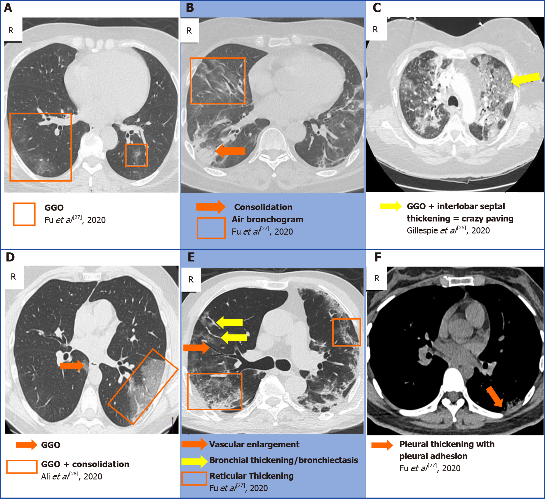
Figure 4 A collection of chest computed tomography that displays some of the classical findings of coronavirus disease 2019 pneumonia[26-28].
A: Ground-glass opacity (GGO); B: Consolidation and air bronchogram; C: Crazy paving; D: GGO superimposed with consolidation; E: Bronchiectasis, reticular thickening, with vascular enlargement; F: Pleural adhesion. A, B, E and F: Citation: Fu Z, Tang N, Chen Y, Ma L, Wei Y, Lu Y, Ye K, Liu H, Tang F, Huang G, Yang Y, Xu F. CT features of COVID-19 patients with two consecutive negative RT-PCR tests after treatment. Science Report 2020; 10: 11548. Copyright ©The Author(s) 2020. Published by Springer Nature; C: Citation: Gillespie M, Flannery P, Schumann JA, Dincher N, Mills R, Can A. Crazy-Paving: A Computed Tomographic Finding of Coronavirus Disease 2019. Clinical Practice and Cases in Emergency Medicine 2020; 4: 461-463. Copyright ©The Author(s) 2020. Published by UC Irvine; D: Citation: Ali TF, Tawab MA, ElHariri MA. CT chest of COVID-19 patients: what should a radiologist know? Egyptian Journal of Radiology and Nuclear Medicine 2020; 51: 120. Copyright ©The Author(s) 2020. Published by Springer Nature.
- Citation: Pal A, Ali A, Young TR, Oostenbrink J, Prabhakar A, Prabhakar A, Deacon N, Arnold A, Eltayeb A, Yap C, Young DM, Tang A, Lakshmanan S, Lim YY, Pokarowski M, Kakodkar P. Comprehensive literature review on the radiographic findings, imaging modalities, and the role of radiology in the COVID-19 pandemic. World J Radiol 2021; 13(9): 258-282
- URL: https://www.wjgnet.com/1949-8470/full/v13/i9/258.htm
- DOI: https://dx.doi.org/10.4329/wjr.v13.i9.258


