Copyright
©The Author(s) 2021.
World J Radiol. Aug 28, 2021; 13(8): 243-257
Published online Aug 28, 2021. doi: 10.4329/wjr.v13.i8.243
Published online Aug 28, 2021. doi: 10.4329/wjr.v13.i8.243
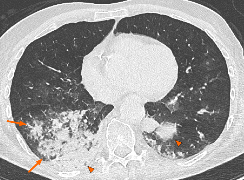
Figure 4 Axial computed tomography image in a 50-year-old woman diagnosed with bronchopneumonia shows confluent centrilobular nodules (arrows) and consolidation (arrowheads) mostly located in the lower lobes.
- Citation: Perrone F, Balbi M, Casartelli C, Buti S, Milanese G, Sverzellati N, Bersanelli M. Differential diagnosis of COVID-19 at the chest computed tomography scan: A review with special focus on cancer patients. World J Radiol 2021; 13(8): 243-257
- URL: https://www.wjgnet.com/1949-8470/full/v13/i8/243.htm
- DOI: https://dx.doi.org/10.4329/wjr.v13.i8.243


