Copyright
©The Author(s) 2021.
World J Radiol. Jun 28, 2021; 13(6): 171-191
Published online Jun 28, 2021. doi: 10.4329/wjr.v13.i6.171
Published online Jun 28, 2021. doi: 10.4329/wjr.v13.i6.171
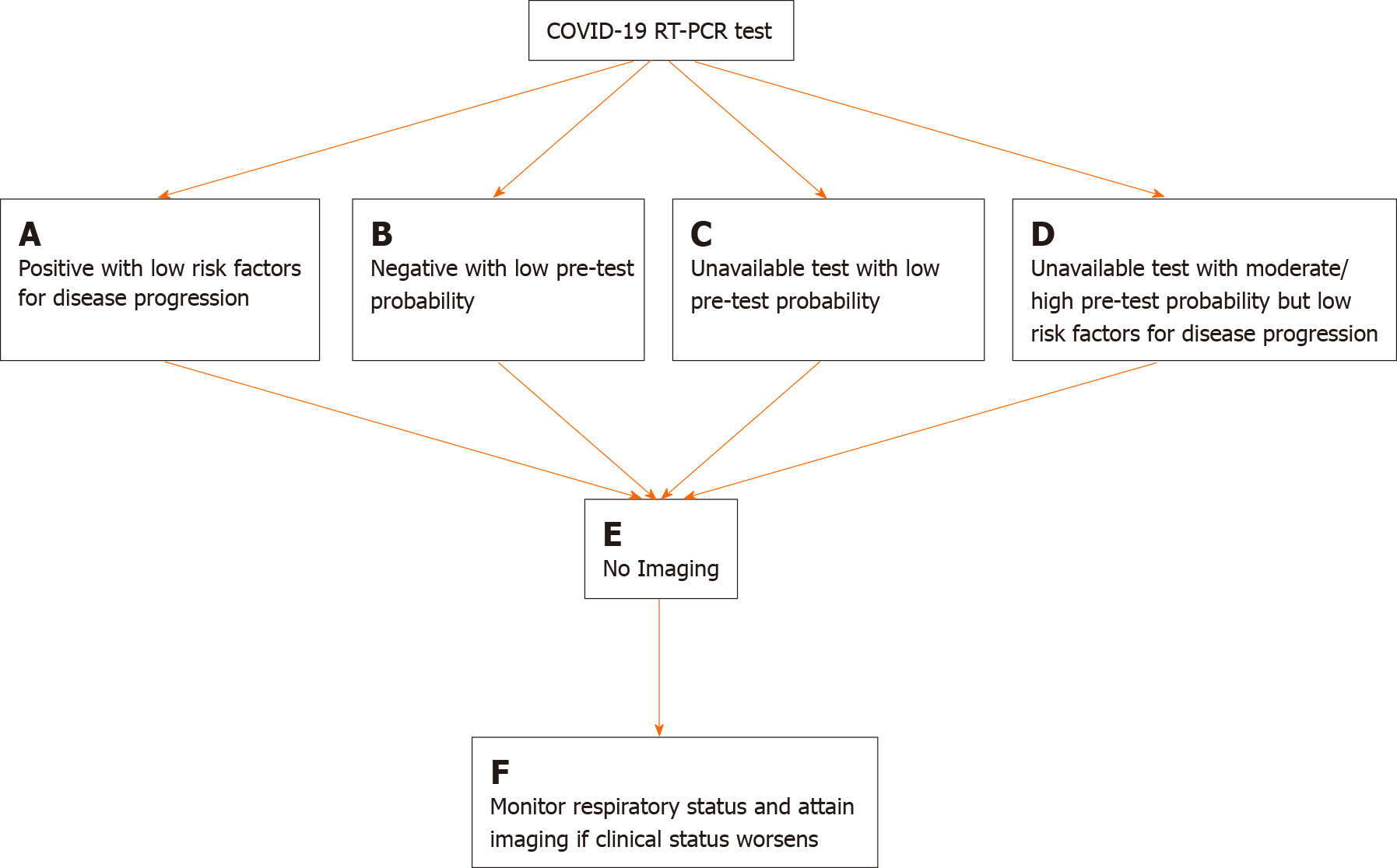
Figure 2 Flowchart depicting four scenarios in which imaging is not indicated in the diagnostic work up and management of coronavirus disease-2019.
A: Low risk factors for disease progression are defined as the absence of underlying comorbidities such as cardiovascular disease, diabetes, hypertension, and an immunocompromised status; B: Low pre-test probability is defined as a low background prevalence of disease in the surrounding area and an unlikely scenario of exposure to severe acute respiratory syndrome coronavirus 2 (SARS-CoV-2); C: Low pre-test probability is defined as a low background prevalence of disease in the surrounding area and an unlikely scenario of exposure to SARS-CoV-2; D: Moderate/high pre-test probability is defined as a high background prevalence of disease in the surrounding area and a likely scenario of exposure to SARS-CoV-2; E: Imaging refers to either the use of chest radiography (CXR) or chest computed tomography (CT). The employment of either is dependent upon time of presentation (early = chest CT, late = CXR), resources (CT scanner availability), and clinical expertise (preference of physician for a particular imaging modality); F: Imaging would ideally be chest radiography as it allows for rapid assessment of an evolving clinical status. This flow chart was adapted and modified based on the Fleischner Society’s article from April of 2020 (Rubin). COVID-19: Coronavirus disease-2019; RT-PCR: Real time-polymerase chain reaction.
- Citation: Pezzutti DL, Wadhwa V, Makary MS. COVID-19 imaging: Diagnostic approaches, challenges, and evolving advances. World J Radiol 2021; 13(6): 171-191
- URL: https://www.wjgnet.com/1949-8470/full/v13/i6/171.htm
- DOI: https://dx.doi.org/10.4329/wjr.v13.i6.171


