Copyright
©The Author(s) 2021.
World J Radiol. Jun 28, 2021; 13(6): 157-170
Published online Jun 28, 2021. doi: 10.4329/wjr.v13.i6.157
Published online Jun 28, 2021. doi: 10.4329/wjr.v13.i6.157
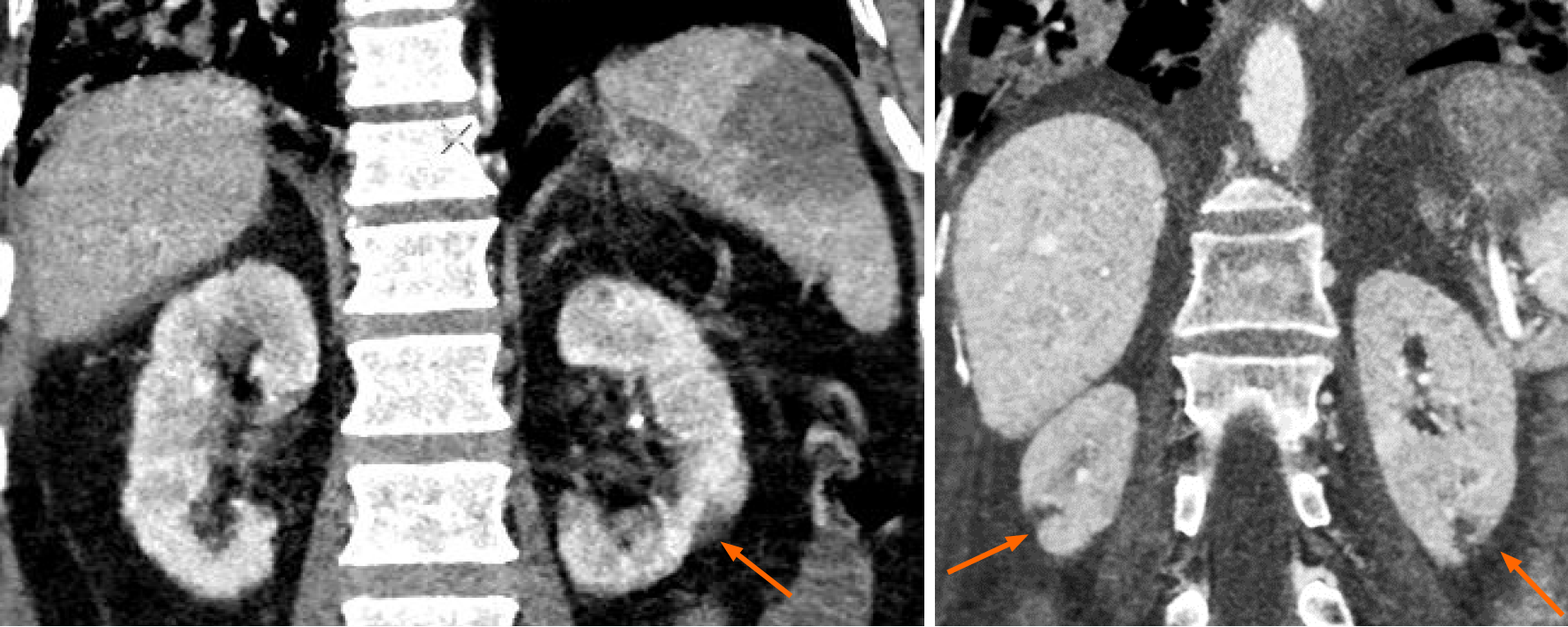
Figure 6 Solid organ infarction in a patient with coronavirus disease 2019.
Coronal contrast enhanced computed tomography (CT) images of the abdomen show discrete, wedge-shaped non-enhancing renal parenchymal defects, with their apex pointing towards the medulla and base parallel to the subcapsular region (orange arrows), suggestive of renal infarcts. These were incidentally detected during CT pulmonary angiography.
- Citation: Vaidya T, Nanivadekar A, Patel R. Imaging spectrum of abdominal manifestations of COVID-19. World J Radiol 2021; 13(6): 157-170
- URL: https://www.wjgnet.com/1949-8470/full/v13/i6/157.htm
- DOI: https://dx.doi.org/10.4329/wjr.v13.i6.157


