Copyright
©The Author(s) 2021.
World J Radiol. Feb 28, 2021; 13(2): 40-52
Published online Feb 28, 2021. doi: 10.4329/wjr.v13.i2.40
Published online Feb 28, 2021. doi: 10.4329/wjr.v13.i2.40
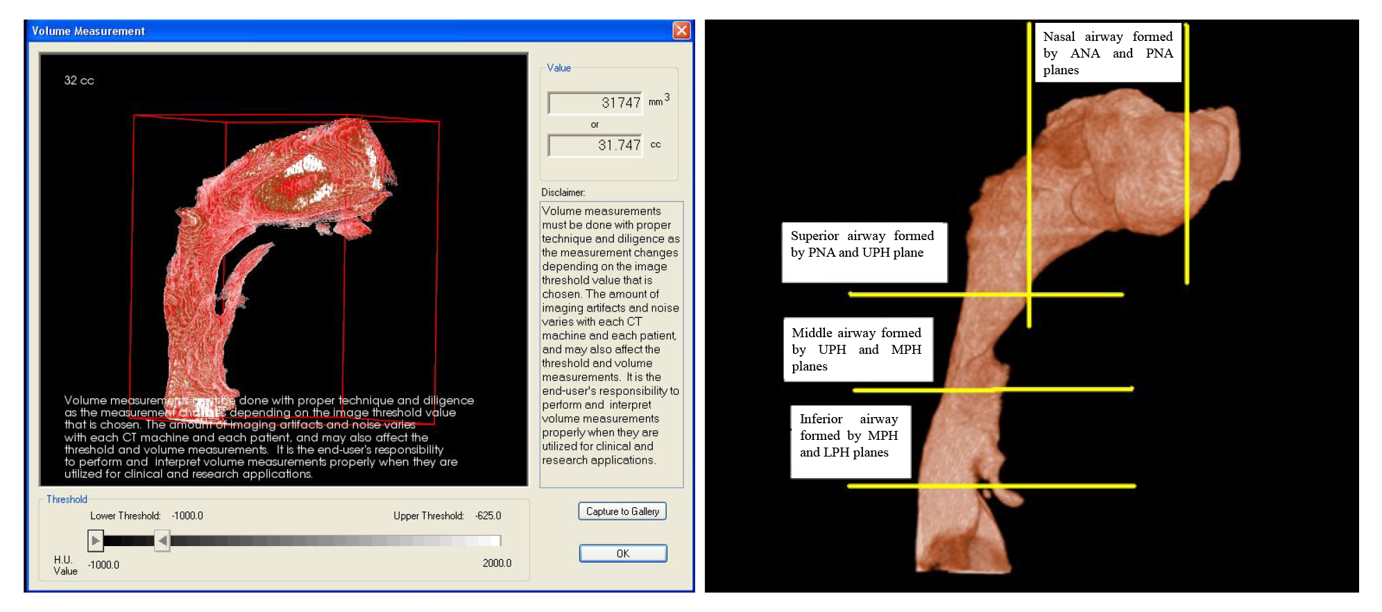
Figure 3 Airway isolated with the software and various referencing plane.
ANA: Anterior nasal; PNA: Posterior nasal; UPH: Upper pharyngeal; MPH: Middle pharyngeal; LPH: Lower pharyngeal.
- Citation: Kochhar AS, Sidhu MS, Bhasin R, Kochhar GK, Dadlani H, Sandhu J, Virk B. Cone beam computed tomographic evaluation of pharyngeal airway in North Indian children with different skeletal patterns. World J Radiol 2021; 13(2): 40-52
- URL: https://www.wjgnet.com/1949-8470/full/v13/i2/40.htm
- DOI: https://dx.doi.org/10.4329/wjr.v13.i2.40


