Copyright
©The Author(s) 2021.
World J Radiol. Jan 28, 2021; 13(1): 29-39
Published online Jan 28, 2021. doi: 10.4329/wjr.v13.i1.29
Published online Jan 28, 2021. doi: 10.4329/wjr.v13.i1.29
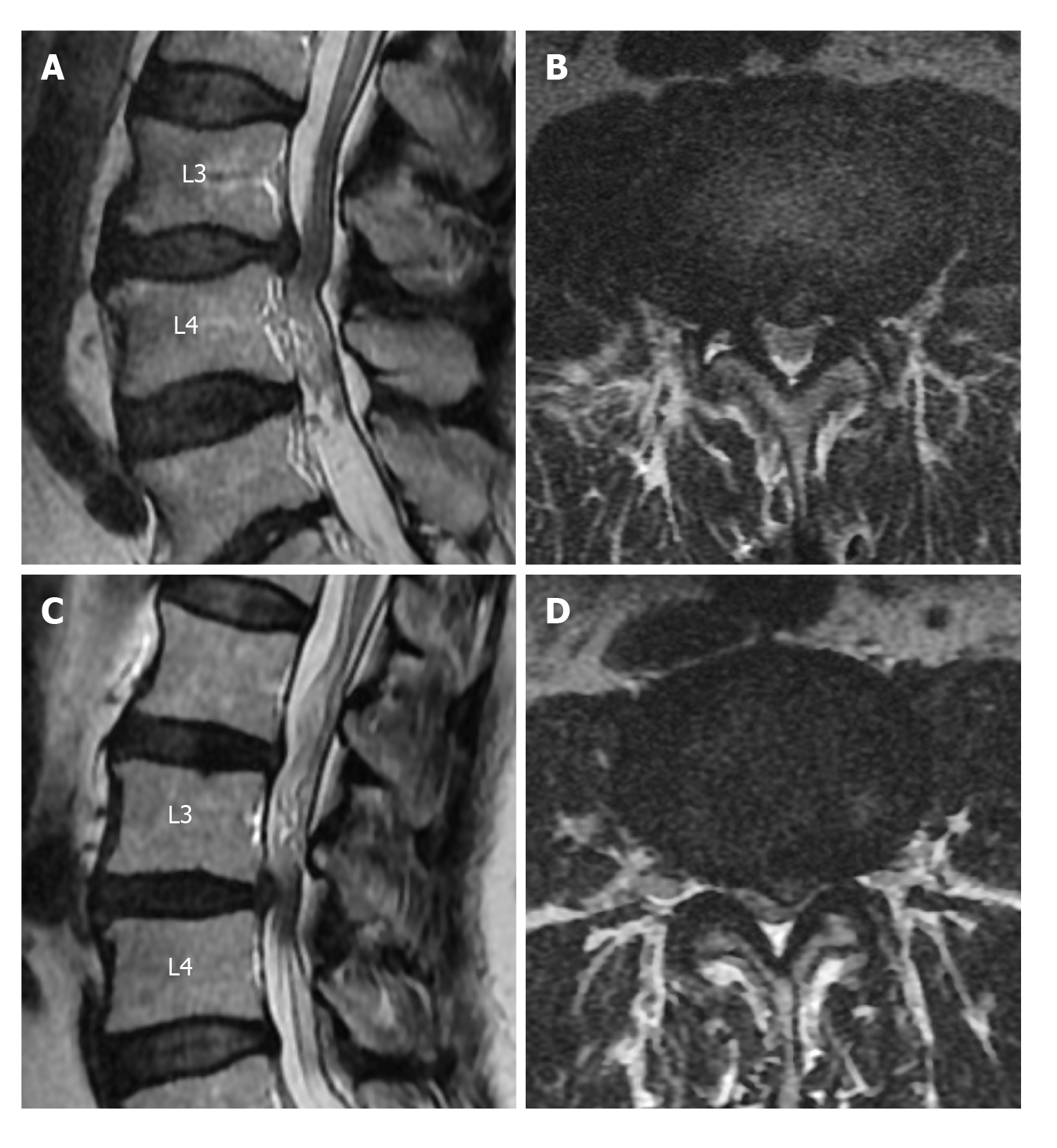
Figure 4 Soft and sharp margin types of disc herniation into the dural sac.
A: On the sagittal T2-weighted image, soft margin disc herniation at the level of L3-L4 intervertebral disc space and redundant nerve roots at the inferior of the stenosis are shown; B: The axial T2-weighted images of soft margin disc herniation are shown; C: On the sagittal T2-weighted image, sharp margin disc herniation at the L3-L4 intervertebral disc space and redundant nerve roots at its superior are shown; D: Axial T2-weighted image of sharp disc herniation is shown.
- Citation: Gökçe E, Beyhan M. Magnetic resonance imaging findings of redundant nerve roots of the cauda equina. World J Radiol 2021; 13(1): 29-39
- URL: https://www.wjgnet.com/1949-8470/full/v13/i1/29.htm
- DOI: https://dx.doi.org/10.4329/wjr.v13.i1.29


