Copyright
©The Author(s) 2021.
World J Radiol. Jan 28, 2021; 13(1): 19-28
Published online Jan 28, 2021. doi: 10.4329/wjr.v13.i1.19
Published online Jan 28, 2021. doi: 10.4329/wjr.v13.i1.19
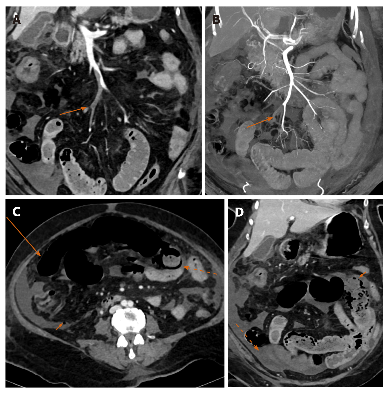
Figure 4 A 65-year-old female with acute mesenteric ischemia.
A: Coronal reformatted image of contrast-enhanced computed tomography (CECT) abdomen shows filling defects (orange arrow) in ileal branches of superior mesenteric artery suggestive of thrombosis; B: Coronal reformatted image of CECT abdomen shows occlusion of accompanying tributaries of superior mesenteric vein (SMV) with superior extension of thrombus into the main stem of SMV; C: Axial CECT image showing dilated small bowel with paper thin wall (long orange arrow), circumferential pneumatosis (dotted orange arrow) and foci of free extraluminal air (small orange arrow) indicating transmural bowel necrosis with perforation; D: Coronal CECT image showing a bowel segment with absent mural enhancement (solid orange arrow), and ascites (dotted arrow).
- Citation: Shankar A, Varadan B, Ethiraj D, Sudarsanam H, Hakeem AR, Kalyanasundaram S. Systemic arterio-venous thrombosis in COVID-19: A pictorial review. World J Radiol 2021; 13(1): 19-28
- URL: https://www.wjgnet.com/1949-8470/full/v13/i1/19.htm
- DOI: https://dx.doi.org/10.4329/wjr.v13.i1.19


