Copyright
©The Author(s) 2021.
World J Radiol. Jan 28, 2021; 13(1): 19-28
Published online Jan 28, 2021. doi: 10.4329/wjr.v13.i1.19
Published online Jan 28, 2021. doi: 10.4329/wjr.v13.i1.19
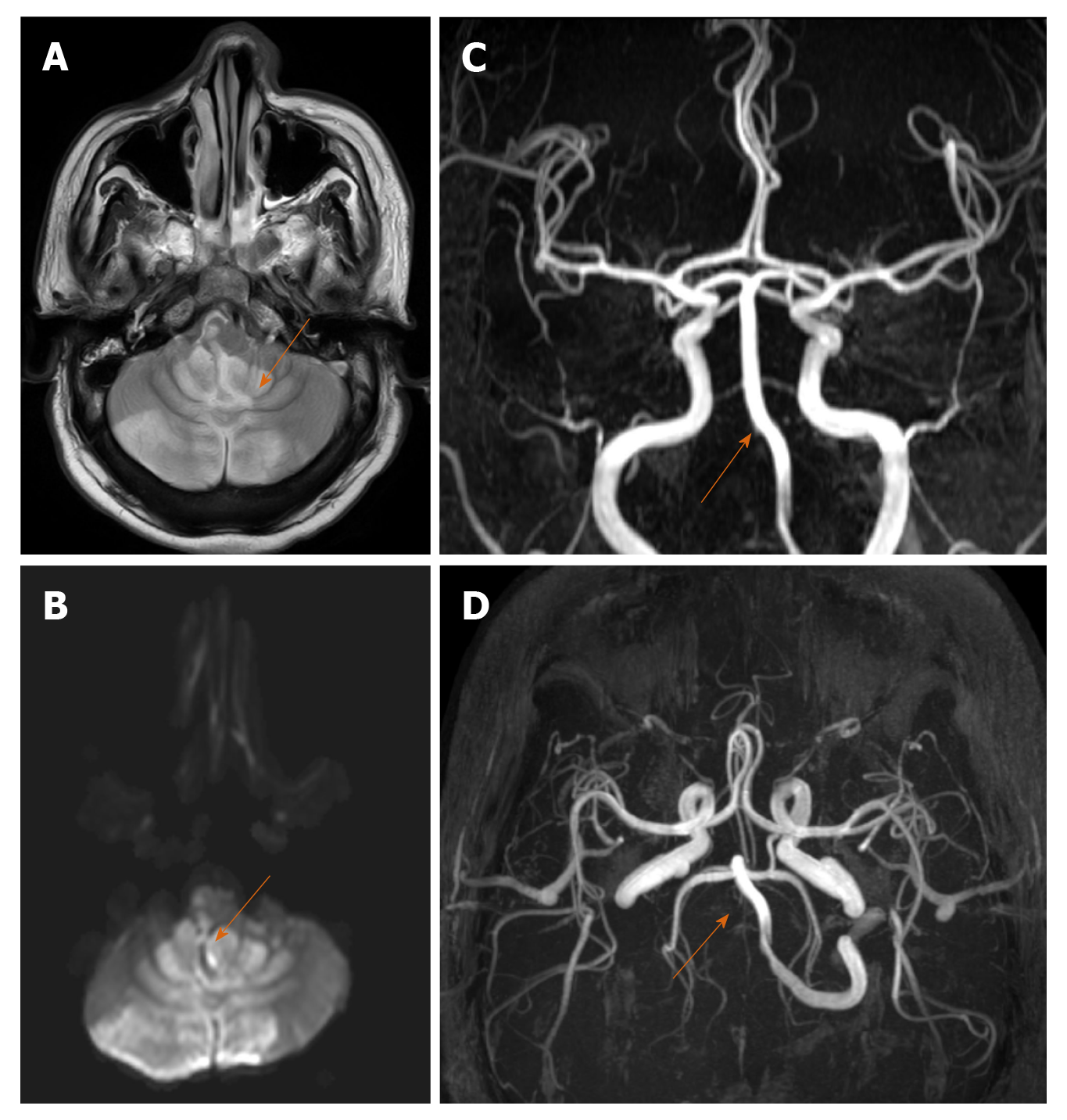
Figure 1 A 35-year-old male with posterior circulation stroke.
A and B: Axial sections of magnetic resonance imaging brain (T2W, diffusion sequences) show areas of high signal in both cerebellar hemispheres, vermis and brainstem suggestive of acute infarcts; C and D: Magnetic resonance artery coronal and axial sections show complete non visualization of right vertebral artery (arrow) suggestive of thrombosis.
- Citation: Shankar A, Varadan B, Ethiraj D, Sudarsanam H, Hakeem AR, Kalyanasundaram S. Systemic arterio-venous thrombosis in COVID-19: A pictorial review. World J Radiol 2021; 13(1): 19-28
- URL: https://www.wjgnet.com/1949-8470/full/v13/i1/19.htm
- DOI: https://dx.doi.org/10.4329/wjr.v13.i1.19


