Copyright
©The Author(s) 2020.
World J Radiol. Sep 28, 2020; 12(9): 195-203
Published online Sep 28, 2020. doi: 10.4329/wjr.v12.i9.195
Published online Sep 28, 2020. doi: 10.4329/wjr.v12.i9.195
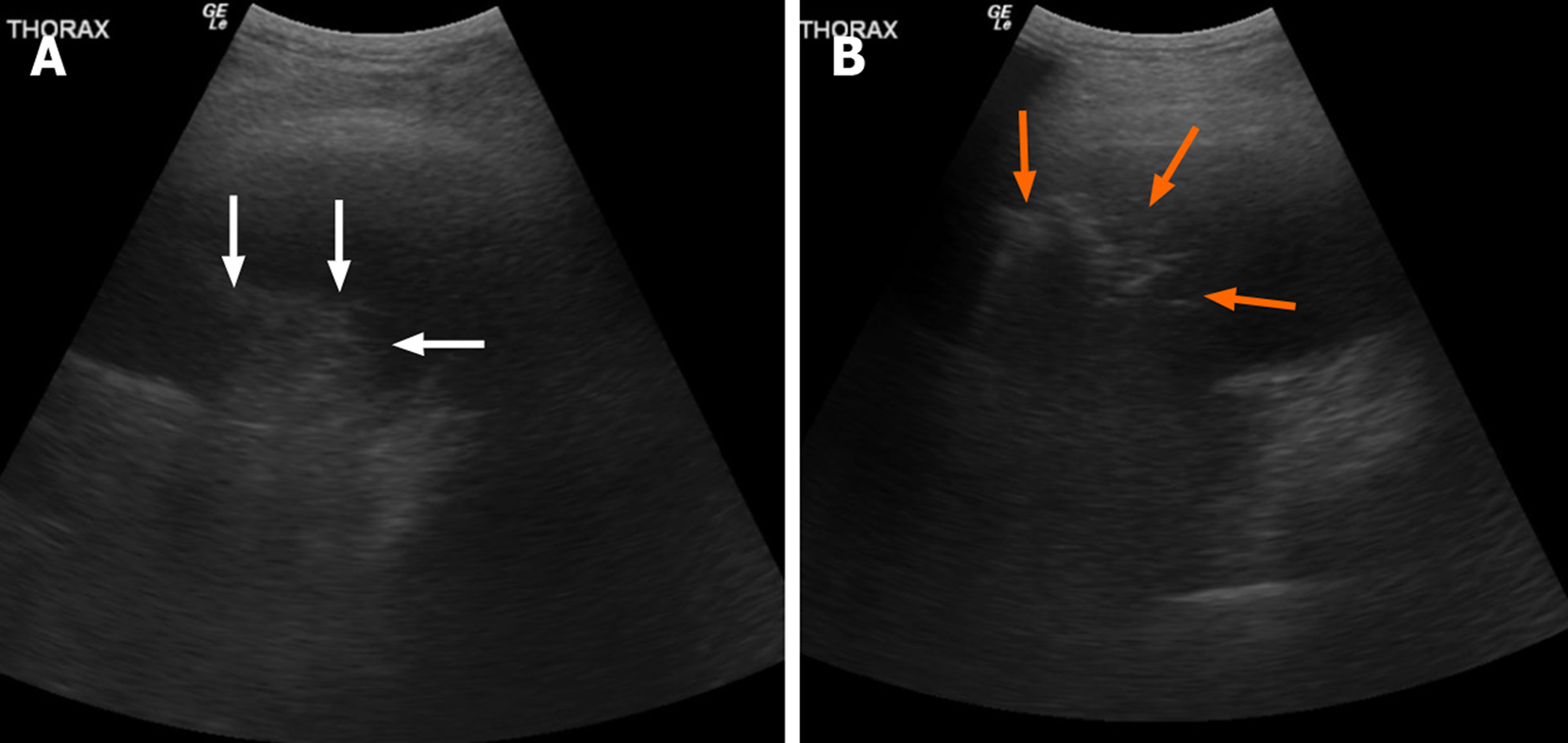
Figure 8 Ultrasonography imaging example of “hepatic pattern” or ”tissue pattern” of the consolidated lung (white arrows) with associated pleural effusion in right lung base (A).
Another example of ultrasonography “shred sign” of pneumonia (orange arrows) with adjacent pleural effusion in left lung base (B).
- Citation: Gandhi D, Ahuja K, Grover H, Sharma P, Solanki S, Gupta N, Patel L. Review of X-ray and computed tomography scan findings with a promising role of point of care ultrasound in COVID-19 pandemic. World J Radiol 2020; 12(9): 195-203
- URL: https://www.wjgnet.com/1949-8470/full/v12/i9/195.htm
- DOI: https://dx.doi.org/10.4329/wjr.v12.i9.195


